


Services on Demand
Journal
Article
 English (pdf)
 Article in xml format
 Article references
 Send this article by e-mail
Indicators
-
 Cited by SciELO
 -
 Access statistics
Related links
-
 Similars in
SciELO
Similars in
SciELO
Share

 Permalink
Medicina Interna
Print version ISSN 0872-671X
Medicina Interna vol.27 no.2 Lisboa Apr. 2020
https://doi.org/10.24950/Imagem/4/20/2/2020
IMAGENS EM MEDICINA / IMAGES IN MEDICINE
Massive Acute Tension Pneumocephalus
Pneumócefalo Hipertensivo Agudo Exuberante
Sónia Canadas  https://orcid.org/0000-0001-8584-3661
Rita Fernandes https://orcid.org/0000-0002-2963-6753
Joana Caires https://orcid.org/0000-0001-5489-0764
Ivan Antunes https://orcid.org/0000-0002-7248-8362
Serviço de Medicina Interna, Hospital Sousa Martins, Guarda, Portugal.
Keywords: Brain Injuries; Pneumocephalus.
Palavras-chave: Lesões Encefálicas; Pneumocéfalo.
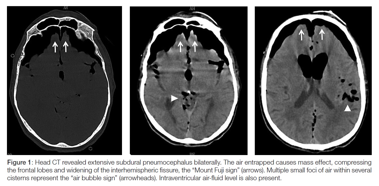
An 86-year-old man was admitted in the emergency room for depressed mental status. He had a Glasgow coma scale (GCS) of 11 points (E4V2M5) and a wound in his frontal region. No other neurological signs were found neither any active haemorrhage. He had normal vital signs. The circumstances were unclear, but he apparently was attacked by his mule. Head computed tomography (CT) showed a large extra-axial pneumocephalus, with signs of tension pneumocephalus (TP) (Fig. 1). The patient was immediately transferred to Neurosurgery (inter-hospital transfer), but due to his comorbidities a conservative approach was rendered with continuous oxygen for 5 days, leading to reduction of the intracranial air. The patient developed fever and was treated with antibiotics and antipyretics, but he never improved to a GCS of 15 points and he died after three weeks.
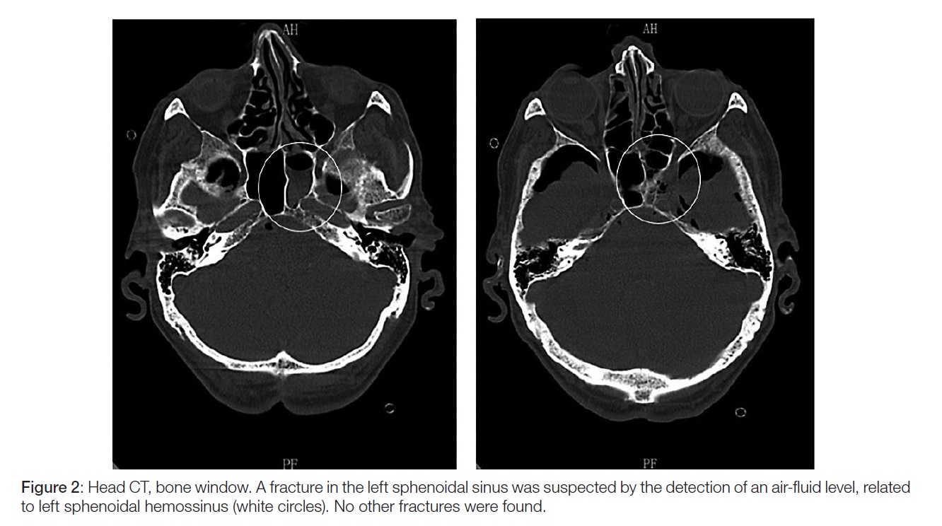
Pneumocephalus occurs when air is present within the intracranial cavity and commonly it is a complication of surgery, trauma, infection or neoplasms.1-3 TP develops when intracranial air causes mass effect on the brain,1-4 and the classic “Mount Fuji sign” and the “air bubble sign” are vital to the diagnosis and to differentiate simple from TP. Pneumocephalus may be seen in 7%-9% of head injuries, and although the precise incidence of TP in those is not known with certainty, a recent review had pointed out to be less than 1%.3 Thereby traumatic TP is rare and is mostly associated with severe craniofacial fractures,2,3 as opposed to our patient (Fig. 2). We considered the publication of this images because the identification of this entity is of the utmost importance to a timely acknowledgment of a serious neurosurgical emergency.
REFERENCES
1. Schirmer CM, Heilman CB, Bhardwaj A. Pneumocephalus: case illustrations and review. Neurocrit Care. 2010;13:152-8. doi: 10.1007/s12028-010-9363-0. [ Links ]
2. Cunqueiro A, Scheinfeld MH. Causes of pneumocephalus and when to be concerned about it. Emerg Radiol. 2018;25:331-40. doi: 10.1007/s10140-018-1595-x. [ Links ]
3. Pillai P, Sharma R, MacKenzie L, Reilly EF, Beery PR 2nd, Papadimos TJ, et al. Traumatic tension pneumocephalus – Two cases and comprehensive review of literature. Int J Crit Illn Inj Sci. 2017;7:58-64. doi: 10.4103/IJCIIS.IJCIIS_8_17.
4. Clement AR, Palaniappan D, Panigrahi RK. Tension pneumocephalus. Anesthesiology. 2017;127:710. doi: 10.1097/ALN.0000000000001703. [ Links ]
Responsabilidades Éticas
Conflitos de Interesse: Os autores declaram a inexistência de conflitos de interesse na realização do presente trabalho.
Fontes de Financiamento: Não existiram fontes externas de financiamento para a realização deste artigo.
Confidencialidade dos Dados: Os autores declaram ter seguido os protocolos da sua instituição acerca da publicação dos dados de doentes.
Proteção de Pessoas e Animais: Os autores declaram que os procedimentos seguidos estavam de acordo com os regulamentos estabelecidos pelos responsáveis da Comissão de Investigação Clínica e Ética e de acordo com a Declaração de Helsínquia da Associação Médica Mundial.
Proveniência e Revisão por Pares: Não comissionado; revisão externa por pares.
Ethical Disclosures
Conflicts of interest: The authors have no conflicts of interest to declare.
Financing Support: This work has not received any contribution, grant or scholarship
Confidentiality of Data: The authors declare that they have followed the protocols of their work center on the publication of data from patients.
Protection of Human and Animal Subjects: The authors declare that the procedures followed were in accordance with the regulations of the relevant clinical research ethics committee and with those of the Code of Ethics of the World Medical Association (Declaration of Helsinki).
Provenance and Peer Review: Not commissioned; externally peer reviewed.
© Autor (es) (ou seu (s) empregador (es)) 2019. Reutilização permitida de acordo com CC BY-NC. Nenhuma reutilização comercial.
© Author(s) (or their employer(s)) 2019. Re-use permitted under CC BY-NC. No commercial re-use.
Correspondence / Correspondência:
Sónia Canadas – soniacanadas@hotmail.com
Serviço de Medicina Interna, Hospital Sousa Martins, Guarda, Portugal
Av. Rainha Dona Amélia, 6300-858 Guarda
Received / Recebido: 16/01/2020
Accepted / Aceite: 05/02/2020
Publicado / Published: 27 de Junho de 2020

