SciELO - Scientific Electronic Library Online

 
vol.28 número3Megacolon TóxicoDoentes Crónicos e Cuidados Paliativos: Da Identificação Precoce ao Cuidado Centrado na Família, num Serviço de Medicina Interna índice de autoresíndice de assuntosPesquisa de artigos
Home Pagelista alfabética de periódicos  

Serviços Personalizados

Journal

Artigo

Indicadores

Links relacionados

  • Não possue artigos similaresSimilares em SciELO

Compartilhar


Medicina Interna

versão impressa ISSN 0872-671X

Medicina Interna vol.28 no.3 Lisboa set. 2021  Epub 01-Dez-2021

https://doi.org/10.24950/cc/93/21/3/2021 

Imagens em Medicina

Osler´S Nodes and Janeway Lesions: From Clinic to Diagnosis

Nódulos de Osler e Lesões de Janeway: Da Clínica ao Diagnóstico

1Serviço de Medicina Interna, Hospital Pedro Hispano, Unidade Local de Saúde de Matosinhos, Matosinhos, Portugal.


A 58-year-old man, with medical background of mitral valve prolapse and dental caries presented with a 12-day evolution of fever, malaise, low back pain and decreased visual acuity. Physical examination revealed small tender purplish nodules on palms of both hands (Fig. 1) and macular hemorrhagic lesions on the right foot (Fig. 2). No recent dental treatments were reported. Complementary study showed mitral valve endocarditis, lumbar spondylodiscitis and retinitis. A methicillin-resistant Staphylococcus aureus was isolated from blood cultures. The patient developed mitral regurgitation due to tendinous cord’s rupture, requiring urgent valve replacement. Valvular vegetations revealed the same agent. Antibiotic treatment with vancomycin, and posteriorly linezolid, led to re-solution of the lesions and clinical improvement.

Figure 1: Osler Nodes (black arrows) on both hands. 

Figure 2: Janeway lesions (black arrows) located on the sole of the left foot. 

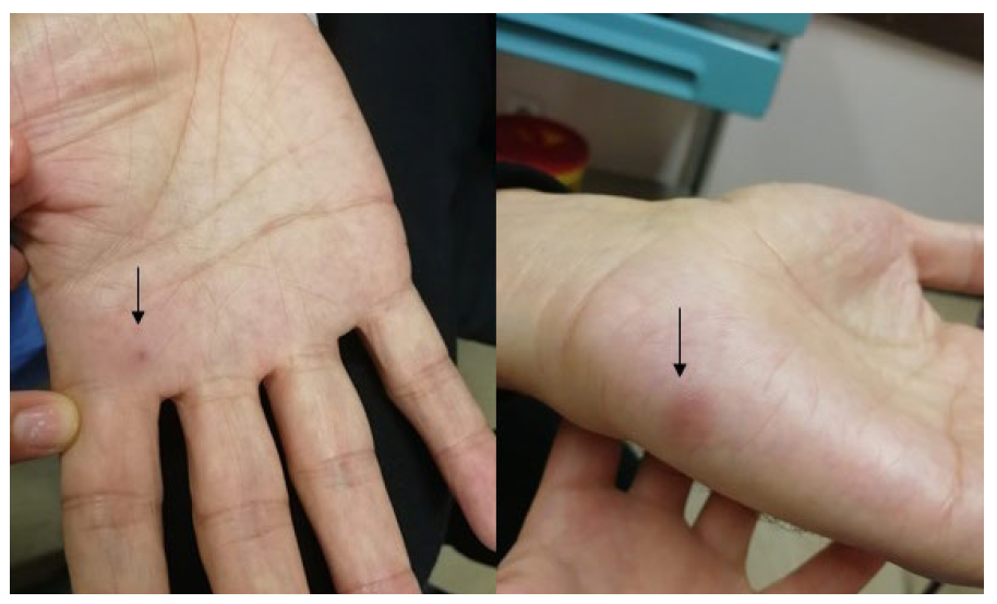
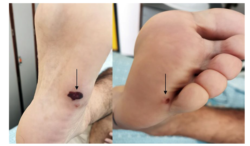
Osler’s nodes are areas of painful nodular erythema, sometimes with a pink colour, usually localized on the tip of the fingers and toes, the sides of the fingers, and the thenar and hypothenar eminences.1Janeway lesions occur on the palms and soles, are non-painful and usually hemorrhagic.1 Infective endocarditis is the most common condition associated with these lesions.1 Their pathogenesis is a contested topic in the literature.1-3 Histological examination of Osler’s nodes often shows findings of sterile vasculitis, so they are considered mostly an immunologic phenomenon.2,3 On the other hand, because histological studies of Janeway lesions have revealed dermal neutrophilic microabscesses without evidence of vasculitis, and bacterial cultures are often positive, these lesions are thought to result directly from septic microemboli.2 Some data revealed that patients with Osler´s nodes and Janeway lesions had a higher rate of infective endocarditis related extracardiac complications,4 which is the case of this patient.

The authors highlight the differences between these two phenomena and their relevance on the diagnosis of infective endocarditis.

REFERÊNCIAS

1. Marrie TJ. Osler's nodes and Janeway lesions. Am J Med. 2008; 121:105-6. doi:10.1016/j.amjmed.2007.07.035 [ Links ]

2. Sethi K, Buckley J, de Wolff J. Splinter haemorrhages, Osler's nodes, Ja-neway lesions and Roth spots: the peripheral stigmata of endocarditis. Br J Hosp Med. 2013;74:C139-42. doi:10.12968/hmed.2013.74.sup9.c139 [ Links ]

1Responsabilidades Éticas Conflitos de Interesse: Os autores declaram a inexistência de conflitos de interesse na realização do presente trabalho. Fontes de Financiamento: Não existiram fontes externas de financiamento para a realização deste artigo. Confidencialidade dos Dados: Os autores declaram ter seguido os protocolos da sua instituição acerca da publicação dos dados de doentes. Consentimento: Consentimento do doente para publicação obtido. Proveniência e Revisão por Pares: Não comissionado; revisão externa por pares. Ethical Disclosures Conflicts of interest: The authors have no conflicts of interest to declare. Financing Support: This work has not received any contribution, grant or scholarship Confidentiality of Data: The authors declare that they have followed the pro-tocols of their work center on the publication of data from patients. Patient Consent: Consent for publication was obtained. Provenance and Peer Review: Not commissioned; externally peer re-viewed

2© Autor (es) (ou seu (s) empregador (es)) e Revista SPMI 2021. Reutiliza-ção permitida de acordo com CC BY-NC. Nenhuma reutilização comercial. © Author(s) (or their employer(s)) and SPMI Journal 2021. Re-use permit-ted under CC BY-NC. No commercial re-use

Received: February 26, 2021; Accepted: March 16, 2021

Correspondence / Correspondência: Ana Catarina Trigo - katarina_alimd@hotmail.com Serviço de Medicina Interna, Hospital Pedro Hispano, Unidade Local de Saúde de Matosinhos, Portugal. Rua Dr. Eduardo Torres, 4464-513 Senhora da Hora

Creative Commons License This is an open-access article distributed under the terms of the Creative Commons Attribution License