SciELO - Scientific Electronic Library Online

 
vol.29 número4Trombocitopenia Imune: Uma Manifestação Hematológica Rara de SarcoidoseEcografia Point-of-Care como Processo Dinâmico no Diagnóstico de Oligoanúria índice de autoresíndice de assuntosPesquisa de artigos
Home Pagelista alfabética de periódicos  

Serviços Personalizados

Journal

Artigo

Indicadores

Links relacionados

  • Não possue artigos similaresSimilares em SciELO

Compartilhar


Medicina Interna

versão impressa ISSN 0872-671X

Medicina Interna vol.29 no.4 Lisboa dez. 2022  Epub 02-Jan-2023

https://doi.org/10.24950/rspmi.634 

Imagens em Medicina/ Images in Medicine

Papillary Fibroelastoma on the Pulmonary Valve: An Uncommon and Unexpected Diagnosis

Fibroelastoma Papilar da Válvula Pulmonar: Um Diagnóstico Raro e Inesperado

Catarina Lameiras1 
http://orcid.org/0000-0001-8302-3037

Inês Fialho2 

Marta Mendes Lopes1 

Maria do Céu Dória1 

1Serviço de Medicina Interna, Hospital Prof. Doutor Fernando da Fonseca, Amadora, Portugal

2Serviço de Cardiologia, Hospital Prof. Doutor Fernando da Fonseca, Amadora, Portugal


A 71-year-old woman was assessed for fatigue in a 20 m flat walk in the previous year. No alterations were found on physical examination. Blood tests were normal, including inflammation parameters, brain natriuretic peptide, thyroid function, and arterial blood gases. Transthoracic echocardiogram revealed preserved left ventricular systolic function, mild right atrial dilatation, mild right ventricular enlargement, elevated pulmonary artery (PA) systolic pressure (55 mmHg), and a large, rounded mass (24x12 mm) apparently adherent to the wall of the main PA, immediately distal to the pulmonary valve, mobile and non-obstructive (Fig. 1).

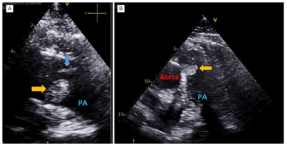
Figure 1: Transthoracic echocardiogram - parasternal short-axis view of the great vessels (A) and subcostal short-axis view (B) - show a rounded well delimitated homogenous mass (24x12 mm) (yellow arrow) apparently adherent to the medial wall of the main pulmonary artery, immediately distal to the pulmonary valve (blue arrow), mobile and non-obstructive . 

Computed tomography angiography confirmed the PA mass (Fig. 2) and was suggestive of distal pulmonary thromboembolism (PTE).

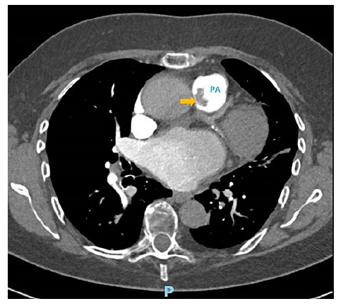
Figure 2: Computed tomography pulmonary angiography show a pulmonary artery mass (yellow arrow) without contrast enhancement or invasion of the surrounding structures. 

Thus, the differential diagnosis comprised a thrombus or a tumor. Cardiac magnetic resonance and body positronemission tomography scan evidenced late gadolinium enhancement of the mass, suggestive of a tumor, without 18-FDG uptake, less suggestive of malignancy. No other lesions were found. Given the dimension and location of the mass and diagnostic uncertainty, the team decided for surgical excision. A gelatinous friable mass with a pedicle attached to the pulmonary valve was removed. The histological exam revealed a papillary fibroelastoma (PF). PF on the pulmonary valve is rare, with only 7% of cases described on this location.1 Pulmonary hypertension (PH) was admitted as a microembolization consequence of direct tumor embolization or possible thrombi formation within the fronds of the PF. An accurate differential diagnosis in the presence of a PA mass is crucial. An incorrect PTE diagnosis, the most frequent diagnosis in this setting, results in anticoagulant therapy initiation with potential adverse effects.2 The differential diagnosis between thrombus and intracardiac tumor depends on an integrated multimodality radiologic study.3,4Furthermore, the presence of PH in association with a PA mass should alert to the possibility of tumor embolization, an uncommon cause of group 4 PH.5

REFERÊNCIAS

1. Gowda RM, Khan IA, Nair CK, Mehta NJ, Vasavada BC, Sacchi TJ. Cardiac papillary fibroelastoma: a comprehensive analysis of 725 cases. Am Heart J. 2003;146:404-10. doi: 10.1016/S0002-8703(03)00249-7 [ Links ]

2. Rudkovskaia AA, Bandyopadhyay D. Intraluminal arterial filling defects misdiagnosed as pulmonary emboli. Clin Chest Med. 2018;39:505-13. doi:10.1016/j.ccm.2018.04.004 [ Links ]

3. Palaskas N, Thompson K, Gladish G, Agha AM, Hassan S, Iliescu C, et al. Evaluation and Management of Cardiac Tumors. Curr Treat Options Cardio-vasc Med. 2018;20:29. doi: 10.1007/s11936-018-0625-z. [ Links ]

4. Xi XY, Gao W, Gong JN, Guo XJ, Wu JY, Yang YH, et al. Value of 18F-FDG PET/CT in differentiating malignancy of pulmonary artery from pulmonary thromboembolism: a cohort study and literature review. Int J Cardiovasc Imaging. 2019;35:1395-403. doi: 10.1007/s10554-019-01553-5. [ Links ]

5. Konstantinides SV, Meyer G, Becattini C, Bueno H, Geersing GJ, et al; ESC Scientific Document Group. 2019 ESC Guidelines for the diagnosis and ma-nagement of acute pulmonary embolism developed in collaboration with the European Respiratory Society (ERS). Eur Heart J. 2020;41:543-603. doi: 10.1093/eurheartj/ehz405 [ Links ]

Fontes de Financiamento: Não existiram fontes externas de financiamento para a realização deste artigo.

4Confidencialidade dos Dados: Os autores declaram ter seguido os protocolos da sua instituição acerca da publicação dos dados de doentes.

5Consentimento: Consentimento do doente para publicação obtido.

6Proveniência e Revisão por Pares: Não comissionado; revisão externa por pares.

Financing Support: This work has not received any contribution, grant or scholarship.

10Confidentiality of Data: The authors declare that they have followed the protocols of their work center on the publication of data from patients.

11Patient Consent: Consent for publication was obtained.

12Provenance and Peer Review: Not commissioned; externally peer re-viewed.

13© Autor (es) (ou seu (s) empregador (es)) e Revista SPMI 2022. Reutiliza-ção permitida de acordo com CC BY-NC. Nenhuma reutilização comercial. © Author(s) (or their employer(s)) and SPMI Journal 2022. Re-use permit-ted under CC BY-NC. No commercial re-use

Received: May 18, 2022; Accepted: July 23, 2022

Correspondence / Correspondência: Catarina Lameiras- anaclameirasm01@gmail.com Serviço de Medicina Interna, Hospital Prof. Doutor Fernando da Fonseca, Amadora, Portugal IC19, 2720-276 - Amadora, Portugal

Declaração de Contribuição CL, IF, MML, MCD - Elaboração e revisão do manuscrito Todos os autores aprovaram a versão final a ser submetida. Contributorship Statement CL, IF, MML, MCD - Preparation and revision of the manuscript All authors approved the final draft.

Conflitos de Interesse: Os autores declaram a inexistência de conflitos de interesse na realização do presente trabalho.

Conflicts of Interest: The authors have no conflicts of interest to declare.

Creative Commons License This is an open-access article distributed under the terms of the Creative Commons Attribution License