


Serviços Personalizados
Journal

Artigo
 Inglês (pdf)
 Artigo em XML
 Referências do artigo
 Enviar este artigo por email
Indicadores
 Citado por SciELO
 Links relacionados
 Similares em
SciELO
Similares em
SciELO Compartilhar

 Permalink
Jornal Português de Gastrenterologia
versão impressa ISSN 0872-8178
J Port Gastrenterol. vol.19 no.1 Lisboa jan. 2012
Splenic nodules in a patient with liver cirrhosis
Nódulos esplénicos em doente com cirrose hepática
Inês Marques*, Ana Lagos, Beatriz Rodrigues, Beatriz Costa Neves
Serviço de Gastrenterologia – Hospital Pulido Valente
*Autor para correspondência
A 72-year-old woman with liver cirrhosis due to hepatitis B was admitted for new onset ascites. On physical examination, her skin and conjunctivas were pale and her abdomen was distended. The spleen was 12 cm palpable and there was no jaundice or lymphadenopathy.
On laboratory investigations, leukopenia (2.7 × 10⁹/L), thrombocytopenia (34.0 × 10⁹/L) and iron deficiency anemia were detected. Her serum albumin, aspartate aminotransferase and alanine aminotransferase levels were 2.2 g/dL, 29 U/L and 20 U/L, respectively. The International normalized ratio was 1.7. Diagnostic paracentesis was performed, which showed a serum-ascites albumin gradient (SAAG) > 1.1, with negative culture. Abdominal fluid cytology revealed white-cell count of 102 cells/µl, with 78% mononuclear cells and was negative for malignant cells.
Abdominal ultrasound was performed and revealed characteristic findings of liver cirrhosis and massive splenomegaly with multiple tiny hyperechoic spots without acoustic shadowing (figure 1). Computed tomographic scan showed multiple faint hypoattenuating lesions in the spleen (figure 2). Magnetic resonance imaging in both T1- and T2-weight sequences showed multiple hypointense, scattered nodules with a diameter of only a few millimeters within the spleen (figure 3), regarded as siderotic nodules (Gamna-Gandy bodies).
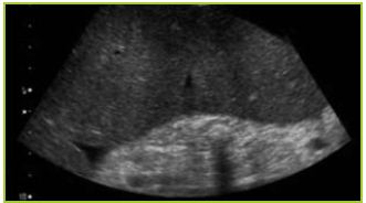
Figure 1 Ultrasound findings: splenomegaly with multiple tiny hyperechoic spots without acoustic shadowing.
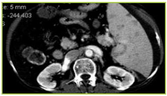
Figure 2 Computed tomographic scan showed multiple faint hypoattenuating lesions in the spleen (arterial fase).
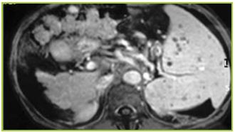
Figure 3 Magnetic resonance imaging (T2-weight sequences) showed multiple hypointense, scattered splenic nodules.
Gamna-Gandy bodies, were firstly described by Marini, in 1902, but this entity became actually known with the studies developed in 1905 and 1921 by Charles Gandy and Carlo Gamna1. They are mainly found in patients with portal hypertension and represent foci of iron deposits resulting from intraparenchymal congestion and hemorrhage.2,3
Ultrasound and computed tomography findings may suggest diagnosis; however, magnetic resonance imaging is the most sensitive imaging modality for detection of these lesions4 Although hypointense on typical MRI pulse sequences (T1, T2), gradient echo sequences are particularly sensitive with rather sharp signal reduction (signal void) related to the susceptibility effects of hemoglobin degradation products3. This appearance is both very sensitive for detection and specific for characterization of these lesions.
Gamna-Gandy bodies must therefore be considered in differential diagnosis of multiple splenic nodules in a patient with long standing portal hypertension.
REFERENCES
1. Unsal N, Erden A, Erden I. Evaluation of the splenic vein diameter and longitudinal size of the spleen in patients with Gamna-Gandy bodies. Diagn Interv Radiol 2006;12:125-128. [ Links ]
2. Selçuk D, Demirel K, Kantarci F, et al. Gamna-Gandy bodies: A sign of portal hypertension. Turk J Gastroenterol 2005;16:150-152. [ Links ]
3. Halefoglu A. Case series: Gamna-Gandy bodies of the spleen: A supportive finding for portal hypertension. Indian J Radiol Imaging 2007;17:186-187. [ Links ]
4. Dobritz M, Nomayr A, Bautz W, et al. Gamna-Gandy bodies detected with MR imaging: a case report. Magn Reson Imaging 2001;19:1249-1251. [ Links ]
*Autor para correspondência
Serviço de Gastrenterologia – Hospital Pulido Valente
Morada: Alameda das Linhas de Torres, 117 – 1000 Lisboa
E-mail: inesnmarques@zonmail.pt
Recebido para publicação: 09/10/2010 e Aceite para publicação: 15/12/2010
