


Serviços Personalizados
Journal
Artigo
 Inglês (pdf)
 Artigo em XML
 Referências do artigo
 Enviar este artigo por email
Indicadores
-
 Citado por SciELO
 -
 Acessos
Links relacionados
-
 Similares em
SciELO
Similares em
SciELO
Compartilhar

 Permalink
Jornal Português de Gastrenterologia
versão impressa ISSN 0872-8178
J Port Gastrenterol. vol.20 no.6 Lisboa dez. 2013
https://doi.org/10.1016/j.jpg.2013.09.003
ENDOSCOPIC SPOT
Esophageal melanocytosis: Case report and literature review
Melanocitose esofágica: relato de caso e revisão da Literatura
Luiz Bertgesa,b,∗, Angelo Macedoc, Moisés Pedrosaa,b, Elise Carvalhoa,b, Gabriela Toledoa, Denise Bittencourta, Gibran Nascifa,b, Carolina Fariaa
a Faculdade de Ciência Médicas e da Saúde de Juiz de Fora - FCMS/JF, Juiz de Fora, MG, Brazil
b Hospital Therezinha de Jesus (HTJ), Juiz de Fora, Brazil
c Santa Casa de Misericórdia de Piumhi-MG, Piumhi, MG, Brazil
*Corresponding author
Introduction
Esophageal melanocytosis is a rare benign entity, with little specificity in terms of symptoms, usually located in the middle and lower thirds of the esophagus, characterized by melanocytic proliferation in the esophageal squamous epithelium and melanin deposition in the mucosa.2-4 Little is known about the etiology and natural course of this condition, although it is hypothesized that it may result from a chronic irritant stimuli such as gastroesophageal reflux disease, chronic esophagitis, which would cause mucosal damage and subsequent reactive melanocytic hyperplasia.2,3,5 This article aims to report a rare case of melanocytosis in a patient with atypical chest pain and dyspepsia, and to review the literature. The evolution of the patient was monitored and a record of new clinical, laboratory, and radiological findings was made, as well as a comparison with other cases reported in the relevant literature.
Case report
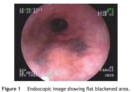
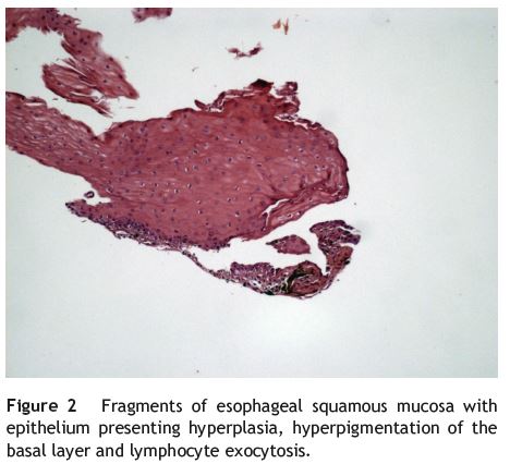
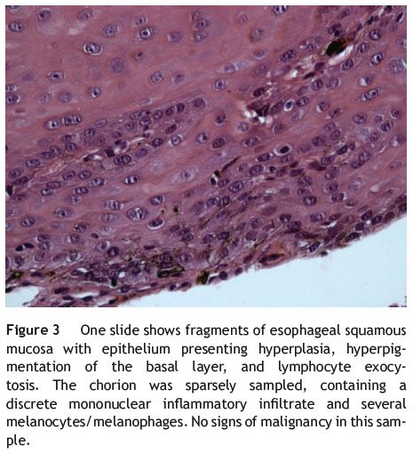
A female patient, aged 45, presented with atypical chest pain and dyspepsia. During upper digestive endoscopy, a flat blackened area was located beginning at 32 cm from the upper dental arch (Fig. 1). The lesion was about 30mm in extent and occupied about 30% of the esophageal circumference, having an interspersed area of mucosa of normal color. Microscopy showed a fragment of esophageal squamous mucosa with the epithelium presenting hyperplasia, hyperpigmentation of the basal layer, and lymphocyte exocytosis (Fig. 2); the chorion was sparsely sampled, containing a discrete mononuclear inflammatory infiltrate and several melanocytes, melanophages with no signs of malignancy (Fig. 3).
Discussion
Esophageal melanocytosis is endoscopically characterized by a circular, linear, or oval lesion of dark-brown color, smooth surface, and jagged edges.4 In histological examination, it is characterized by melanocytic proliferation in the esophageal squamous epithelium and by mucosal melanin deposition.4,5 Proliferation of melanocytes is seldom observed, with an estimated incidence of about 0.07-0.15%.5 Differential diagnoses should include melanocytic nevi and malignant melanoma.4 We can differentiate melanocytosis from malignant melanoma by the absence of spindle cells and cytologic atypia in the histopathology exam, and endoscopically the melanoma assumes a polypoid form.1,4 Anthracosis, hemosiderosis, dye intake, and lipofuscin deposition should also be considered as differential diagnoses.4
References
1. Álvarez R, Funke R, Solis F, Molina H, Pacheco F, Farias H. Melanoma primario del esófago. Rev Chilena Cir. 2009;61(2):168-70. [ Links ]
2. Cardoso C, Freire R, Gamito E, Quintana C, Cremers I, Oliveira AP. Esophageal melanocytosis. J Port Gastrenterol. 2012;19(3):158-9. [ Links ]
3. Chang F, Deere H. Esophageal melanocytosis morphologic features and review of the literature. Arch Pathol Lab Med. 2006;130(4):552-7. [ Links ]
4. Denız K, Çelıkbılek M, Torun E, Yücesoy M. Turk J Gastroenterol. 2010;21(3):321-2. [ Links ]
5. Wang DG, Li XG, Gao H, Sun XY, Zhou XQ. Coexistence of esophageal blue nevus, hair follicles and basaloid squamous carcinoma: a case report. World J Gastroenterol. 2008;14(26):4253-6. [ Links ]
Ethical disclosures
Protection of human and animal subjects. The authors declare that no experiments were performed on humans or animals for this study.
Confidentiality of data. The authors declare that they have followed the protocols of their work center on the publication of patient data and that all the patients included in the study received sufficient information and gave their written informed consent to participate in the study.
Right to privacy and informed consent. The authors have obtained the written informed consent of the patients or subjects mentioned in the article. The corresponding author is in possession of this document.
Conflicts of interest
The authors have no conflicts of interest to declare.
*Corresponding author
Correio eletrónico: denisebittencourt@live.com (L. Bertges).
Received 4 February 2013; accepted 3 September 2013
