SciELO - Scientific Electronic Library Online

 
 número13Instrumentos de educação alimentar e nutricional para crianças em idade pré-escolar: necessidades e desafios atuais índice de autoresíndice de assuntosPesquisa de artigos
Home Pagelista alfabética de periódicos  

Serviços Personalizados

Journal

Artigo

Indicadores

Links relacionados

  • Não possue artigos similaresSimilares em SciELO

Compartilhar


Millenium - Journal of Education, Technologies, and Health

versão impressa ISSN 0873-3015versão On-line ISSN 1647-662X

Mill  no.13 Viseu dez. 2020  Epub 31-Dez-2020

https://doi.org/10.29352/mill0213.09.00334 

Education and social development

Dynamic changes of the pelvic floor in elite athletes of different sports.

Alterações dinâmicas do pavimento pélvico em atletas de elite de diferentes desportos.

Cambios dinámicos del suelo pélvico en atletas de élite de diferentes deportes.

1 Universidade de Trás-os-Montes e Alto Douro, Departamento de Ciências do Desporto, Exercício e Saúde, Vila Real, Portugal.

2 Universidade de Trás-os-Montes e Alto Douro, Escola Superior de Saúde, Vila Real, Portugal.

3 Universidade de Trás-os-Montes e Alto Douro, Departamento de Ciências do Desporto, Exercício e Saúde, CIDESD, CITAB, Vila Real, Portugal

4 Universidade de Trás-os-Montes e Alto Douro, Departamento de Ciências do Desporto, Exercício e Saúde, CITAB, Vila Real, Portugal

5 Centro Hospitalar de Trás-os-Montes e Alto Douro, Departamento de Obstetrícia e Ginecologia, Vila Real, Portugal

6 Universidade Fernando Pessoa, Faculdade Ciências da Saúde, Escola Superior de Saúde, Porto, Portugal

7 Hospital São João, Porto, Portugal


Abstract

Introduction:

One of the functions of the pelvic floor muscles (PFM) is to support the pelvic organs and continence. This continence mechanism tends to change when PFM are exposed to high-impact exercises.

Objetives:

To describe the dynamic changes in the pelvic floor (PF) in elite nulliparous athletes.

Methods:

Translabial two and three-dimensional ultrasound was used to assess PF anatomy and function in athletes (n=8). This ultrasonography was performed after voiding and in the supine position, using a vaginal probe. The descent of the pelvic organs was assessed on a maximum Valsalva maneuver, whilst the volume datasets were acquired at rest, during maximum voluntary contraction (MVC) and during a Valsalva maneuver. The athletes performed each maneuver at least 3 times, with the most effective being used for evaluation.

Results:

The bladder neck descent was 14 mm for the javelin thrower, being the highest value when compared to the remaining participants. Three athletes featured the rectocele (swimming, gymnastics and javelin throw) and 4 participants presented a paravaginal defect (volleyball, horsemanship, javelin throw and printer). The volleyball athlete had the highest value of the levator hiatal area in MVC value.

Conclusions:

The athletes present minimal differences in the evaluated parameters. The sample is small to generalize the results, but there is a tendency for athletes of high-impact exercises to have a lower CMV value. Further studies are needed to corroborate these results.

Keywords: maximal voluntary contraction; translabial ultrasound; levator ani; pelvic floor function

Resumo

Introdução:

Uma das funções dos músculos do pavimento pélvico (MPP) é suportar os órgãos pélvicos e a continência. Este mecanismo da continência tende a alterar-se quando os MPP estão expostos a exercícios de alto-impacto.

Objetivos:

Descrever as alterações dinâmicas no pavimento pélvico (PP) em atletas de elite, nulíparas.

Métodos:

Foi utilizada a ecografia translabial para avaliar a anatomia e a função do PP nas atletas (n=8), tendo sido realizada após a micção e em decúbito dorsal através de sonda vaginal. A descida dos órgãos pélvicos foi avaliada em manobra de Valsalva. O volume foi avaliado em repouso, durante a contração máxima voluntária (CMV) e em Valsalva. As atletas realizaram cada manobra pelo menos 3 vezes, sendo a mais eficaz utilizada para avaliação.

Resultados:

A descida do colo vesical foi de 14 mm para a atleta de lançamento de dardo, sendo o valor mais alto quando comparado com as restantes participantes. Três atletas apresentaram retocele (natação, ginástica e lançamento de dardo) e 4 participantes apresentaram defeito paravaginal (voleibol, equitação, lançamento de dardo e corrida). A voleibolista apresentou o maior valor da área hiatal do músculo elevador durante a CMV.

Conclusões:

As atletas mostraram diferenças mínimas nos parâmetros avaliados. A amostra foi pequena para generalizar resultados, mas há uma tendência para as atletas de exercícios de alto impacto apresentarem a menor CMV. São necessários mais estudos para corroborar estes resultados.

Palavras-chave: contração máxima voluntária; ecografia translabial; elevador do ânus; função do pavimento pélvico

Resumen

Introducción:

Una de las funciones de los músculos del suelo pélvico (MSP) es apoyar los órganos pélvicos y la continencia. Este mecanismo de continencia tiende a cambiar cuando las MSP están expuestas a ejercicios de alto impacto.

Objetivos:

Describir los cambios dinámicos en el suelo pélvico (SP) en atletas nulíparos de élite.

Métodos:

Se utilizó ultrasonido translabial para evaluar la anatomía y la función de la SP en atletas (n = 8), que se realizó después de orinar y en posición supina a través de un tubo vaginal. El descenso de los órganos pélvicos se evaluó mediante la maniobra de Valsalva. El volumen se evaluó en reposo, durante la contracción voluntaria máxima (CVM) y en Valsalva. Los atletas realizaron cada maniobra al menos 3 veces, siendo la más efectiva la utilizada para la evaluación.

Resultados:

El descenso del cuello de la vejiga fue de 14 mm para el atleta de jabalina, el valor más alto en comparación con los otros participantes. Tres atletas presentaron rectocele (natación, gimnasia y jabalina) y 4 participantes presentaron defectos paravaginales (voleibol, equitación, jabalina y carrera). El jugador de voleibol presentó el valor más alto del área hiatal del músculo elevador durante el CVM.

Conclusiones:

Los atletas mostraron diferencias mínimas en los parámetros evaluados. La muestra fue pequeña para generalizar los resultados, pero los atletas de ejercicios de alto impacto tienden a tener el CVM más bajo. Se necesitan más estudios para corroborar estos resultados.

Palabras clave: contracción voluntaria máxima; ultrasonido translabial; elevador ano; función del suelo pélvico

Introduction

The World Health Organization considers urinary incontinence to be a public health problem that mostly affects women and consequently compromises their quality of life. Some people still see this pathology as a natural consequence of age and adapt to the changes imposed by it. Nowadays it is known that it is not just a natural consequence of age, as it can also appear in young people, including nulliparous women and athletes.

1. Theoretical framework

The important role of the pelvic floor muscles (PFM) is to support the pelvic organs, continence and childbirth (Karim, Begum et al. 2019). Continence is a complex mechanism, involving the coordination between the PFM, the urethra, the bladder and their supporting ligaments. This mechanism involves both striated (voluntary control) and smooth (involuntary) muscles (Bø 2004) The aim of this study was to describe the dynamic changes in the pelvic floor in elite nulliparous athletes.

2. Methods

2.1 Sample

This study, reporting the PF function by translabial 2D/3D ultrasonography in a sample of 8 athletes, volunteered to participate in the study. All athletes were active in the first league of their various sports and at the time of testing had been training for a minimum of 5 years, having reached a national or international level of competition. In this research note, the sociodemographic and anthropometric data were collected using a structured questionnaire. The inclusion criteria were elite nulliparous athletes trained or qualified in a particular sport, between 18 and 30 years old. The exclusion criteria were the inability to perform a correct PFM contraction, combination of multiple sports and surgical treatment of gynecological and urological illnesses.

2.2 Data collection instruments and procedures

The sports included were: volleyball (1), swimming (n=1), karate (n=1), gymnastics (n=1), horsemanship (n=1), pentathlon (n=1), javelin throw (n=1) and sprinting (n=1). Ethical approval was obtained from the Ethics Committee of the Hospital center of Trás-os-Montes and Alto Douro, Vila Real, Portugal (165/2018). All participants gave informed written consent. The evaluation and imaging were performed using GE Voluson 730 pro and were carried out by an experienced doctor, who did not have access to all the clinical information. All athletes were imaged in a supine position, after voiding. Imaging was performed in the mid-sagittal plane with the angle of acquisition set at 90º. The posterior vesico-urethral angle was acquired at rest and during a Valsalva maneuver. The most effective of at least three maneuvers was used for evaluation. The descent of the pelvic organs was assessed during the Valsalva maneuver. The levator hiatus was measured at rest, during the Valsalva maneuver and MVC of PF.

2.3 Statistical analysis

Sample characteristics (age and BMI) were described using mean±standard deviation. For the biometric indices, the value of each athlete is presented.

3. Results

The results from the questionnaire indicate that all were asymptomatic for SUI or gynecological disorders. Eight international female league players, of different sports (age, 22.4±4.1 years and body mass, 20.7±1.9 kg) (Table 1)

Concerning the 2D ultrasound, the findings were the following: the PVU angle measurements were similar, showing no difference between them, either at rest or Valsalva. Only the karate athlete has urethral hypermobility because her value during the Valsalva maneuver was 164.40º, although it was asymptomatic. The results of bladder neck descent, relative to the symphysis pubis on maximum Valsalva demonstrate that the most favorable value was in volleyball, with no movement/descent of the bladder, while the worst value was in the javelin throw (still this value is within normal). Three athletes presented a mild rectocele, javelin throw, gymnastics and swimming modalities (Table 2).

Concerning the 3D ultrasound evaluations: the area of the levator hiatus at rest, the Valsalva and the MVC were measured. At rest, the lowest value was in pentathlon and the highest value was in javelin throw. For the Valsalva maneuver, the javelin throw athlete showed the highest value and the pentathlon obtained the lowest value. Regarding the MVC, the lowest value was in gymnastics and the highest value was in volleyball. As for the LAM assessment, no participant showed LAM avulsion. A paravaginal defect was detected in than half of the sample (n= 4). The athletes with the defect were in volleyball, horsemanship, pentathlon and sprinting (Table 2). There were no changes in normality with respect to the levator hiatal area and LAM avulsion (Figure 1).

Table 1 Participants’ characteristics (n=9) 

Note. BMI, body mass índex; SD, standard deviation

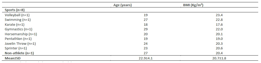
Table 2 Biometric indices of the sui, pop, levator hiatos area, lam avulsion and paravaginal defect 

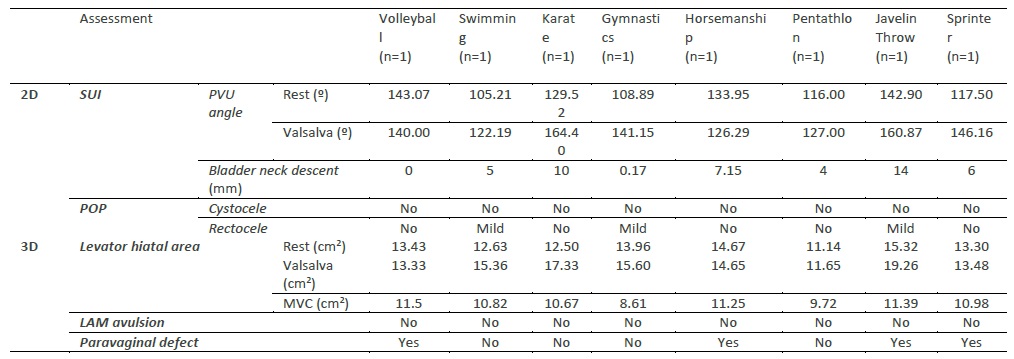
Note. 2D, two-dimensional ultrasound; 3D, three-dimensional ultrasound; LAM, levator ani muscle; MVC, maximum voluntary contraction; POP, pelvic organ prolapse; PVU, posterior vesico-urethral; SUI, stress urinary incontinence.

Figure 1 Measurement of maximum voluntary contraction (A) in the minimal hiatal dimensions. 

4. Discussion

Previous studies using 3D ultrasound imaging had evaluated functional and morphological parameters of young nulliparous athletes, such PF trauma and distension of the levator hiatos (Dietz 2019). Kruger et al (Kruger, Dietz et al. 2007) found the levator hiatal area at rest, 12.71±2.49 cm2 in the high-impact, frequent intense training (HIFIT) group and 12.77±2.43 cm2 in the control group. Found the levator hiatal area on valsalva, 21.53±9.98 cm2 in the HIFIT group and 14.91±7.18 cm2 in the control group. Found the levator hiatal area on pelvic floor muscle contraction, 10.59±1.71 in the HIFIT group and 9.72±2.11 in the control group . These findings are similar to this study. The dynamic assessment of the LAM was able to demonstrate that no participant presented LAM avulsion. Despite this, the javelin throw athlete had a markedly increased levator hiatus area during a voluntary Valsalva maneuver. This might be explained by a higher awareness by high-impact athletes regarding the kinesthetics involved in high impact exercises, which can enable them to use task-specific muscles, as well as more abdominal strength, and the resulting increase in intra-abdominal pressure they are able to develop (Kruger, Dietz et al. 2007). The volleyball athlete had the highest value of the levator hiatal area in MVC, so she contracted the PFM less. As explained above, high-impact exercises can cause PF disorders, namely weakening of the PFM, which in turn can lead to SUI (Carvalhais, Da Roza et al. 2018). This tends to be more prevalent in high-impact sports (Pires, Pires et al. 2020), such as gymnastics, track and field, and some ball games (Gram and Bø 2020).

SUI is mainly related to bladder neck hypermobility, PFM and pelvic ligaments which are weak or damaged (Lasak, Jean-Michel et al. 2018), as well as changes to the structure of the neuromuscular and connective tissues supporting the bladder neck and the urethra. If athletes continue to strengthen their PFM, urine leakage may never happen (Livingston 2016). It is extremely important to have PFM training in these athletes, as it is advised for the prevention and treatment of SUI in several studies (Hagovska, Švihra et al. 2017). In order to better accomplish the MVC, the participants received information about the structure / function of the PFM, in addition to learning to develop awareness of them. This means that the athletes received assistance in contracting the correct muscle during the MVC, without contracting the other related muscles, such as the rectus abdominis, the thigh adductor and the gluteus maximus (accessories). Doing so would mean a considerable decrease in the contractile activity of the PFM (Kruger, Dietz et al. 2007). SUI can interfere with athletes’s participation in sport and fitness activities, consequently affecting their health, self-esteem and well-being (Pires T 2020).

Two-dimensional (2D) ultrasonography has been one of the tools used to assess anatomical changes in the mobility of the pelvic organ of women suffering from SUI and POP (Dietz 2017). The findings showed that the PVU angle at rest does not show major changes. The findings during the Valsalva maneuver, demonstrated one participant (karate) had parameters compatible with urethral hypermobility. Although karate is considered a moderate impact sport, in fact the athlete mentioned that running, a high impact sport (Mitchell, Haskell et al. 2005), is also part of her daily training, which may justify these results. Women with SUI commonly (or high impact sports) show an increase in bladder neck mobility and a descent below the level of the symphysis pubis during the Valsalva maneuver. Dietz et al (Dietz, Eldridge et al. 2004) noted that the HIFIT nulliparous women group showed a considerably higher mobility of the bladder neck, as well as in the levator hiatal area than the control group. However, most of the HIFIT women were asymptomatic for UI, as observed in our study as well. The bladder neck descent measurements were the highest in the javelin throw (even though, within normal ranges). The remaining measurements were within the normal parameters (Dietz 2004). There is no definition of ‘normal’ for bladder neck descent, although a cut-off of 2.5 cm has been proposed to define hypermobility (Dietz 2004). Urethral mobility as a measure of urethral support, focuses mainly on the bladder neck (Dietz 2017). There is minimal information available on the mobility of the remainder of the urethra, which we believe is important to understand urethral support and the pathophysiology of SUI and urodynamic stress incontinence (Dietz 2019). According to Dietz et al (Dietz 2004), the retrovesical (or posterior urethrovesical) angle usually opens to up to 160-180◦, from a normal value of 90-120◦. Such a change in the retrovesical angle is often (but not always) associated with funneling. Three athletes (swimming, gymnastics and javelin throw) demonstrated a mild rectocele, but were asymptomatic for clinical symptoms of POP. The women with increased pelvic organ descent are usually associated to having ‘weak’ PFM. There is a known correlation between hypermobility syndrome and some connective tissue disorders, including an increased risk for POP (Pirpiris, Shek et al. 2010). These differences may be explained by the differences in the connective tissue itself or the muscle biomechanics, which may have originated before or after the high-impact training (Kruger, Dietz et al. 2007).

Conclusions

Although the sample is very small for large conclusions, there is a tendency for high-impact sports to suffer more changes in PFM, namely weak MVC that may lead to the appearance of SUI, as was the case with volleyball and javelin throw athletes.

Future research with a larger sample of high-performance athletes is necessary to explore whether they present similar results after the intervention.

Acknowledgements

This work is supported by National Funds by FCT - Portuguese Foundation for Science and Technology, under the projects UIDB/04033/2020 and UID04045/2020

References

Bø, K., & Borgen, J. S. (2001). Prevalence of stress and urge urinary incontinence in elite athletes and controls. Medicine and Science in Sports and Exercise, 33(11), 1797-1802. DOI: https://doi.org/10.1097/00005768-200111000-00001 [ Links ]

Bø, Kari. (2004a). Urinary incontinence, pelvic floor dysfunction, exercise and sport. Sports Medicine (Auckland, N.Z.), 34(7), 451-464. DOI: https://doi.org/10.2165/00007256-200434070-00004 [ Links ]

Bø, Kari. (2004b). Pelvic floor muscle training is effective in treatment of female stress urinary incontinence, but how does it work? International Urogynecology Journal and Pelvic Floor Dysfunction, 15(2), 76-84. DOI: https://doi.org/10.1007/s00192-004-1125-0 [ Links ]

Brandão, S., Parente, M., Silva, A. R., Roza, T. D., Mascarenhas, T., Ramos, I., & Jorge, R. M. N. (2015). The Impairment of Female Pelvic Ligaments and Its Relation With Pelvic Floor Dysfunction: Biomechanical Analysis. Computational and Experimental Biomedical Sciences: Methods and Applications, 63-73. DOI: https://doi.org/10.1007/978-3-319-15799-3_4 [ Links ]

Cardoso, A. M. B., Lima, C. R. O. de P., & Ferreira, C. W. S. (2018). Prevalence of urinary incontinence in high-impact sports athletes and their association with knowledge, attitude and practice about this dysfunction. European Journal of Sport Science, 18(10), 1405-1412. DOI: https://doi.org/10.1080/17461391.2018.1496146 [ Links ]

Carvalhais, A., Roza, T. D., & Sacomori, C. (2018). Pelvic Floor in Female Athletes: From Function to Dysfunction. Women’s Health and Biomechanics, 145-153. DOI: https://doi.org/10.1007/978-3-319-71574-2_12 [ Links ]

Carvalho, C., da Silva Serrão, P. R. M., Beleza, A. C. S., & Driusso, P. (2020). Pelvic floor dysfunctions in female cheerleaders: A cross-sectional study. International Urogynecology Journal, 31(5), 999-1006. DOI: https://doi.org/10.1007/s00192-019-04074-w [ Links ]

Casey, E. K., & Temme, K. (2017). Pelvic floor muscle function and urinary incontinence in the female athlete. The Physician and Sportsmedicine, 45(4), 399-407. DOI: https://doi.org/10.1080/00913847.2017.1372677 [ Links ]

Dietz, H. P. (2004a). Ultrasound imaging of the pelvic floor. Part I: Two-dimensional aspects. Ultrasound in Obstetrics & Gynecology: The Official Journal of the International Society of Ultrasound in Obstetrics and Gynecology, 23(1), 80-92. DOI: https://doi.org/10.1002/uog.939 [ Links ]

Dietz, H. P. (2004b). Ultrasound imaging of the pelvic floor. Part II: Three-dimensional or volume imaging. Ultrasound in Obstetrics & Gynecology: The Official Journal of the International Society of Ultrasound in Obstetrics and Gynecology, 23(6), 615-625. DOI: https://doi.org/10.1002/uog.1072 [ Links ]

Dietz, H. P., & Bennett, M. J. (2003). The effect of childbirth on pelvic organ mobility. Obstetrics and Gynecology, 102(2), 223-228. DOI: https://doi.org/10.1016/s0029-7844(03)00476-9 [ Links ]

Dietz, H. P., Eldridge, A., Grace, M., & Clarke, B. (2004). Pelvic organ descent in young nulligravid women. American Journal of Obstetrics and Gynecology, 191(1), 95-99. DOI: https://doi.org/10.1016/j.ajog.2004.01.025 [ Links ]

Dietz, H. P., Shek, C., & Clarke, B. (2005). Biometry of the pubovisceral muscle and levator hiatus by three-dimensional pelvic floor ultrasound. Ultrasound in Obstetrics & Gynecology: The Official Journal of the International Society of Ultrasound in Obstetrics and Gynecology, 25(6), 580-585. DOI: https://doi.org/10.1002/uog.1899 [ Links ]

Dietz, Hans Peter. (2011). Pelvic floor ultrasound in prolapse: What’s in it for the surgeon? International Urogynecology Journal, 22(10), 1221-1232. DOI: https://doi.org/10.1007/s00192-011-1459-3 [ Links ]

Dietz, Hans Peter. (2017). Pelvic Floor Ultrasound: A Review. Clinical Obstetrics and Gynecology, 60(1), 58-81. DOI: https://doi.org/10.1097/GRF.0000000000000264 [ Links ]

Dietz, Hans Peter. (2019). Pelvic floor ultrasound: Essentials. Ultrasound in Medicine and Biology, 45, S60. DOI: https://doi.org/10.1016/j.ultrasmedbio.2019.07.610 [ Links ]

Eliasson, K., Larsson, T., & Mattsson, E. (2002). Prevalence of stress incontinence in nulliparous elite trampolinists. Scandinavian Journal of Medicine & Science in Sports, 12(2), 106-110. DOI: https://doi.org/10.1034/j.1600-0838.2002.120207.x [ Links ]

Gao, Y., Zhao, Z., Yang, Y., Zhang, M., Wu, J., & Miao, Y. (2020). Diagnostic value of pelvic floor ultrasonography for diagnosis of pelvic organ prolapse: A systematic review. International Urogynecology Journal, 31(1), 15-33. DOI: https://doi.org/10.1007/s00192-019-04066-w [ Links ]

Gram, M. C. D., & Bø, K. (2020). High level rhythmic gymnasts and urinary incontinence: Prevalence, risk factors, and influence on performance. Scandinavian Journal of Medicine & Science in Sports, 30(1), 159-165. DOI: https://doi.org/10.1111/sms.13548 [ Links ]

Gray, T. G., & Radley, S. C. (2020). Pelvic Organ Prolapse. Urologic Principles and Practice, 487-497. DOI: https://doi.org/10.1007/978-3-030-28599-9_29 [ Links ]

Hagovska, M., Švihra, J., Buková, A., Horbacz, A., Dračková, D., Švihrová, V., & Kraus, L. (2017). Prevalence of Urinary Incontinence in Females Performing High-Impact Exercises. International Journal of Sports Medicine, 38(3), 210-216. DOI: https://doi.org/10.1055/s-0042-123045 [ Links ]

Haylen, B. T., Maher, C. F., Barber, M. D., Camargo, S., Dandolu, V., Digesu, A., Goldman, H. B., Huser, M., Milani, A. L., Moran, P. A., Schaer, G. N., & Withagen, M. I. J. (2016). An International Urogynecological Association (IUGA) / International Continence Society (ICS) joint report on the terminology for female pelvic organ prolapse (POP). International Urogynecology Journal, 27(2), 165-194. DOI: https://doi.org/10.1007/s00192-015-2932-1 [ Links ]

Haylen, B. T., Ridder, D., Freeman, R. M., Swift, S. E., Berghmans, B., Lee, J., Monga, A., Petri, E., Rizk, D. E., Sand, P. K., & Schaer, G. N. (2010). An International Urogynecological Association (IUGA)/International Continence Society (ICS) joint report on the terminology for female pelvic floor dysfunction. Neurourology and Urodynamics, 29(1), 4-20. DOI: https://doi.org/10.1002/nau.20798 [ Links ]

Karim, R., Begum, S., Ayub, S., Pervaiz, K. F., & Akhtar, R. (2019). INCONTINENCE OF URINE IN PREGNANT WOMEN. Journal of Postgraduate Medical Institute (Peshawar - Pakistan), 33(2), Article 2. https://jpmi.org.pk/index.php/jpmi/article/view/2427Links ]

Kisner, C., & Colby, L. A. (2009). Exercícios terapêuticos: Fundamentos e técnicas (5a edição). Manole. [ Links ]

Kruger, J. A., Dietz, H. P., & Murphy, B. A. (2007). Pelvic floor function in elite nulliparous athletes. Ultrasound in Obstetrics & Gynecology: The Official Journal of the International Society of Ultrasound in Obstetrics and Gynecology, 30(1), 81-85. DOI: https://doi.org/10.1002/uog.4027 [ Links ]

Kruger, J. A., Heap, S. W., Murphy, B. A., & Dietz, H. P. (2008). Pelvic floor function in nulliparous women using three-dimensional ultrasound and magnetic resonance imaging. Obstetrics and Gynecology, 111(3), 631-638. DOI: https://doi.org/10.1097/AOG.0b013e3181655dc2 [ Links ]

Lasak, A. M., Jean-Michel, M., Le, P. U., Durgam, R., & Harroche, J. (2018). The Role of Pelvic Floor Muscle Training in the Conservative and Surgical Management of Female Stress Urinary Incontinence: Does the Strength of the Pelvic Floor Muscles Matter? PM & R: The Journal of Injury, Function, and Rehabilitation, 10(11), 1198-1210. DOI: https://doi.org/10.1016/j.pmrj.2018.03.023 [ Links ]

Livingston, B. (2016). Anatomy and Neural Control of the Lower Urinary Tract and Pelvic Floor. Topics in Geriatric Rehabilitation, 32(4), 280-294. DOI: https://doi.org/10.1097/TGR.0000000000000123 [ Links ]

Louis-Charles, K., Biggie, K., Wolfinbarger, A., Wilcox, B., & Kienstra, C. M. (2019). Pelvic Floor Dysfunction in the Female Athlete. Current Sports Medicine Reports, 18(2), 49-52. DOI: https://doi.org/10.1249/JSR.0000000000000563 [ Links ]

Lourenco, T. R. de M., Matsuoka, P. K., Baracat, E. C., & Haddad, J. M. (2018). Urinary incontinence in female athletes: A systematic review. International Urogynecology Journal, 29(12), 1757-1763. DOI: https://doi.org/10.1007/s00192-018-3629-z [ Links ]

Mitchell, J. H., Haskell, W., Snell, P., & Van Camp, S. P. (2005). Task Force 8: Classification of sports. Journal of the American College of Cardiology, 45(8), 1364-1367. DOI: https://doi.org/10.1016/j.jacc.2005.02.015 [ Links ]

Pires, T. F., Pires, P. M., Moreira, M. H., Gabriel, R. E. C. D., João, P. V., Viana, S. A., & Viana, R. A. (2020). Pelvic Floor Muscle Training in Female Athletes: A Randomized Controlled Pilot Study. International Journal of Sports Medicine, 41(4), 264-270. DOI: https://doi.org/10.1055/a-1073-7977 [ Links ]

Pires, T., Pires, P., Moreira, H., Gabriel, R., Viana, S., & Viana, R. (2020). Assessment of pelvic floor muscles in sportswomen: Quality of life and related factors. Physical Therapy in Sport, 43, 151-156. DOI: https://doi.org/10.1016/j.ptsp.2020.02.015 [ Links ]

Pires, T., Pires, P., Moreira, H., & Viana, R. (2020). Prevalence of Urinary Incontinence in High-Impact Sport Athletes: A Systematic Review and Meta-Analysis. Journal of Human Kinetics, 73, 10. [ Links ]

Pires, T., Pires, P., Moreira, M., & Viana, R. (2020). Prevalence of Urinary Incontinence in High-Impact Sport Athletes: A Systematic Review and Meta-Analysis. Journal of Human Kinetics, 73, 279-288. DOI: https://doi.org/10.2478/hukin-2020-0008 [ Links ]

Pirpiris, A., Shek, K. L., & Dietz, H. P. (2010). Urethral mobility and urinary incontinence. Ultrasound in Obstetrics & Gynecology, 36(4), 507-511. DOI: https://doi.org/10.1002/uog.7658 [ Links ]

Teixeira, R. V., Colla, C., Sbruzzi, G., Mallmann, A., & Paiva, L. L. (2018). Prevalence of urinary incontinence in female athletes: A systematic review with meta-analysis. International Urogynecology Journal, 29(12), 1717-1725. DOI: https://doi.org/10.1007/s00192-018-3651-1 [ Links ]

Ying, T., Li, Q., Xu, L., Liu, F., & Hu, B. (2012). Three-dimensional Ultrasound Appearance of Pelvic Floor in Nulliparous Women and Pelvic Organ Prolapse Women. International Journal of Medical Sciences, 9(10), 894-900. DOI: https://doi.org/10.7150/ijms.4829 [ Links ]

Received: April 27, 2020; Accepted: July 17, 2020

Corresponding Author Telma Pires Universidade Trás-os-Montes e Alto Douro Quinta de Prados 5000-801 Vila Real telmafilipapires@gmail.com

Creative Commons License This is an open-access article distributed under the terms of the Creative Commons Attribution License