


 English (pdf)
 Article in xml format
 Article references
 Send this article by e-mail
 Cited by SciELO
  Similars in
SciELO
Similars in
SciELO 
 Permalink
Introduction
Biological profile features, such as age, sex, ancestry, and stature, are key elements in reconstructing human identity. When unidentified remains show an advanced stage of degradation and/or a high degree of destruction, teeth can offer valuable information due to their morphological features and great resistance to extreme environmental conditions.1,2
Dental radiographs obtained through conventional x-rays, such as periapical, bitewing, occlusal, and orthopantomogram (OPG), provide two-dimensional (2D) data of the lower face in clinical settings. These 2D images have also been conventionally used for human identification in forensic contexts.1 Conebeam
computed tomography (CBCT) has started being widely incorporated in dental clinics due to its many advantages and possibilities. This imaging technique can generate three-dimensional (3D) high-resolution oral and maxillofacial images after reconstructing data from axial, sagittal, and coronal planes.3
Different forensic odontology groups proposed using 3D image-based techniques by CBCT to obtain more accurate biological profile features, such as age and sex.4-8Some of those methods include volumetric and dimensional assessment of the dental crown,4,5 determination of the pulp cavity volume,6,7and volumetric analysis of the pulp/tooth ratio.8
Among permanent teeth, mandibular canines are considered those that exhibit the greatest sexual dimorphism and are therefore adopted, by many, as the tooth of choice for sexual estimation studies.4,6,9,10From a methodologic point of view, the mandibular canine index appears in the literature as an adequate and easy predictor for sex estimation.11 However, the mesiodistal width of a mandibular canine crown has recently been suggested as a simpler technique with higher sex prediction accuracy.10
Like the rest of the teeth, canines show resilience to extreme physical and chemical conditions observed in circumstances where human identification is challenging, such as mass disasters, crashes, or fires.12-14 Moreover, they have a long and strong single root inserted in the alveolar bone, which favors its permanence in the oral arch.15 Nevertheless, a significant impact on teeth can lead to crown fragmentation, causing all metric methods using the canine’s crown dimensions to become useless under these circumstances. The development of methods for sex estimation based on teeth roots can therefore be an alternative or complementary tool when other methods are not applicable. Accordingly, this study aims to evaluate the potential of the buccolingual (BL) dimension of the permanent mandibular canine roots as a sex estimator, using sagittal CBCT images.
Material and methods
This retrospective transversal observational study was conducted at a university dental clinic in Portugal. The sample was composed of 58 Portuguese patients (27 female and 31 male) drawn from non-randomly selected patients who attended the dental clinic from January 2019 to January 2020.
The inclusion criteria were: patients aged 18 to 60 years of Portuguese nationality and Portuguese descent presenting at least one intact mandibular canine. The exclusion criteria were: patients showing canines with therapeutic, pathological, or physiological root modification affecting the cervical region (such as cervical caries, restorations or sealants, abrasion or attrition injuries, and rotations) and images of canines without a distinctive cementoenamel junction (CEJ). Patients aged over 60 years were excluded to reduce the number of individuals with missing teeth or morphological alterations, frequently associated with advanced age.
This study followed the ethical standards of the responsible committee on human experimentation (institutional and national) and the Helsinki Declaration of 1975, as revised in 2008. The study was approved by the institution’s Ethics Committee (Protocol number: 4/CE-IUCS/2020). Written informed consent was obtained from all patients.
The buccolingual (BL) length of the permanent mandibular canine’s cervical margin was obtained using CBCT images from the ProMax 3D Plus device (Planmeca, Finland). A voxel size of 0.2 mm and 0.15 mm and the same energy parameters (90 kV and 10 mA) were used. The maximum BL dimension was measured in the midsagittal plane, as shown in Figure 1.
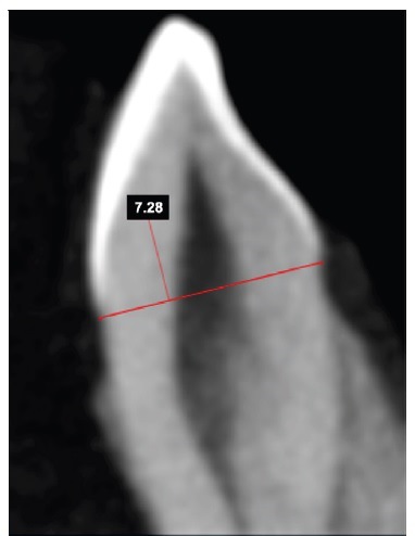
Figura 1 Measurement of the buccolingual root dimension on the cervical margin of a mandibular canine.
On the midsagittal reconstruction, the axial plane that intercepted the most apical extension of the enamel on the buccal or the lingual surface was established as the cervical margin.
The Romexis® 3D imaging software was used to draw straight lines from the CEJ (from buccal to lingual) and measure the distances. A single previously calibrated observer performed the measurements three times within a timeframe of 4 days and was blind to previous measurements. In order to consider cases with a single mandibular canine, left or right mandibular canines were used indiscriminately.
The statistical analysis was performed using the IBM SPSS Statistics for Windows (V.25.0) software program. The Bland-Altman method was performed with the 2nd and 3rd measurements for reliability analysis (Figure 2). The final BL data corresponded to the average of these two measurements.

Figura 2 Bland-Altman analysis related to the buccolingual dimension on the 2nd and 3rd measurements (mm) with the mean error and upper and lower limits for a 95% probability.
The assumption of normality was accepted for the Bland-Altman measures (KS; p> 0.05). The error between the measures was -0.0024 ± 0.029 mm. The intra-observer error was between -0.0612 mm and 0.0564 mm, with a probability of 95%. The error measurement was lower than the difference between the two groups (males and females), which was 0.77 mm. In order to apply the t test for independent samples, the assumption of normality and homogeneity of variances were tested by a Kolmogorov-Smirnov test and a Levene’s test, respectively. The groups were compared using the t test, followed by the accuracy analysis with the receiver operating characteristics (ROC) curve, after calculating the sensitivity and specificity. Then, a cut-off value was obtained discriminating males from females. A statistical significance level of 0.05 was considered.
Results
The male group had an average BL measure (7.03 ± 0.69 mm) higher than the female group (6.26 ± 0.49 mm). The box plot (Figure 3) shows that the measurements taken from a male’s mandibular canine are remarkably larger than those of a female.
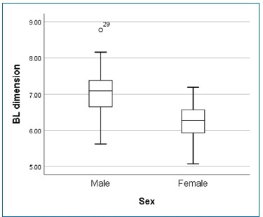
A median of 7.09 mm and an interquartile range of 0.77 mm were obtained for the male sex, and 6.28 mm and 0.78 mm, respectively, for the female sex.
The normality (KS; p>0.05) and the homogeneity of the variances (F=2.524; p>0.12) were accepted for both populations.
The mean difference between sexes in the BL root dimension (0.78±0.16 mm) was statistically significant (t56 df = 4.871; p<0.0005).
The ROC curve is presented in Figure 4. The area under the curve (AUC) was 83.5%, and it was statistically significant (p<0.0005). For an optimal cut-off of 6.64 mm for the BL root dimension, the overall accuracy was around 79%. The percentage of males correctly classified was 77.4%, whereas for females
was 81.5% (Figure 4).

Discussion
Sex estimation methods are fundamental in the process of human identification, and when sex cannot be determined, the reconstruction of the identity is compromised.4,16In the absence of pelvic or skull bones (the gold standards for sex estimation) or when the bone analysis is inconclusive, teeth can be an alternative way to rapidly estimate an individual’s sex.10,17
The odontometric difference between males and females is generally explained by specific gene expression patterns.
Consequently, absolute tooth size and proportions tend to be greater in males.18 Canines are considered the key teeth for sex estimation, showing the highest sexual dimorphism, followed by premolars, first and second molars, and maxillary incisors.4,19 Mandibular canines are the preferred teeth of many researchers aiming to develop sex estimation methods based on tooth dimensions. Most of these methods consider mesiodistal crown distance and/or mandibular inter-canine width.4,9,10,11,20-23Studies measuring teeth root dimensions for sex estimation are much less frequent, although some anthropological works showed positive results using canine root length or neck dimensions by measuring directly on teeth or through x-ray images.24-27
Although canines are very solid teeth, in violent situations such as crash disasters, natural disasters, terrorist attacks, and fires, or simply by prolonged exposure to taphonomic phenomena, teeth crown fragmentation may occur, making methods based on crown measurements impossible. The strong and protected canine root may then serve as an alternative solution. In situations where the dental material is in a deteriorated and fragile state, computed tomography can provide a non-destructive approach and collect not only 3D images but also dental midsagittal planes that are impossible to obtain with conventional x-rays.28 Yet, its accuracy and real forensic application in dental post-mortem evaluation need to be fully demonstrated.
CBCT is a complex technique that also has some limitations: i) CBCT technicians/operators need to be trained; ii) measuring precautions need to be taken to avoid poor image quality (beam hardening, image noise, scatter, ring artifacts, motion artifacts), which could result in the exclusion of many images;29 iii) the CBCT device is expensive and is not available in all dental clinics, legal medicine institutes or scientific police departments. Other limitations in studies like this include pathological or physiological modifications of the teeth (mainly tooth rotation and cervical attrition) that hinder or prevent the image analysis. However, post-mortem computed tomography is being positively evaluated as a complementary method
for dental identification purposes nowadays.30,31Moreover, the costs of acquiring a device are currently decreasing, and new improvements such as the development of mobile units are being carried out.32,33In our view, we are likely to soon witness a widespread use of the CBCT in multiple forensic scenarios.
In this pilot study, we aimed to provide data showing the potential of mandibular canine roots for estimating sex in na adult Portuguese population. A statistically significant difference was found between males and females concerning the cervical BL root dimension of the mandibular canine, and a cut-off was provided. One limitation of this method is the appearance of wear in the cervical region of the canine, more frequent in advanced ages. In these cases, it would not be possible to use the proposed methodology since the original tooth dimension would be altered. Other therapeutic, pathological, or physiological root modifications affecting the cervical region, such as caries, restorations, or rotations, must also be previously checked by the dentist (or trained operator) to avoid misinterpretations. No differences between left and right mandibular canines were found by other authors in a Portuguese population when the mesial-distal dimension of the crown was used as a sex estimator.10 It would be necessary to confirm whether it is also the case for the cervical BL root dimension.
This pilot study used a small non-random sample; hence, further research in a larger and random population is required to fully validate the usefulness of the proposed method. However, we believe that these preliminary results deserve attention and that this method can contribute in the future as a complementary tool for sex estimation in a forensic context.
Conclusion
The cervical buccolingual root dimension on the mandibular canine showed statistically significant differences between sexes in a Portuguese population sample. This dimension may contribute as a complementary tool for sex estimation, particularly when the canine crown is unavailable. Nevertheless, further studies are needed to validate its application in a forensic context.

