SciELO - Scientific Electronic Library Online

 
vol.16 número4Justifica-se a vacinação contra o HPV nas mulheres entre os 26-45 anos?Diagnóstico e abordagem de hyperreactio luteinalis: uma entidade rara na gravidez índice de autoresíndice de materiabúsqueda de artículos
Home Pagelista alfabética de revistas  

Servicios Personalizados

Revista

Articulo

Indicadores

Links relacionados

  • No hay articulos similaresSimilares en SciELO

Compartir


Acta Obstétrica e Ginecológica Portuguesa

versión impresa ISSN 1646-5830

Acta Obstet Ginecol Port vol.16 no.4 Algés dic. 2022  Epub 31-Dic-2022


Case Report/Caso Clínico

Extensive uterine deciduosis as cause for spontaneous hemoperitoneum in pregnancy

Deciduose uterina exuberante como causa de hemoperitoneu espontâneo na gravidez

Diana Margarida Monteiro1 
http://orcid.org/0000-0001-9238-7158

Mafalda Laranjo1 

Inês Sarmento Gonçalves1 

Fátima Soares1 

1. Hospital Pedro Hispano - Unidade Local de Saúde de Matosinhos. Portugal.


Abstract

Ectopic decidualization is usually an incidental finding during surgical approach of a pregnant abdominal cavity. It presents as one or multiple friable and haemorrhagic lesions. We report a pregnant woman presenting with intense abdominal pain at 36 weeks and known placenta previa. During c-section, hemoperitoneum was observed originating on an exuberant friable and highly haemorrhagic lesion, covering the posterior uterine surface, both adnexal regions and the anterior wall of the rectum. Haemostasis was done with conservative measures and the patient had favourable outcome. Deciduosis is a benign entity and should be managed conservatively to prevent iatrogenic lesions.

Keywords: Hemoperitoneum; Pregnancy complications; Decidua

Resumo

A decidualização ectópica é habitualmente um achado incidental durante a intervenção cirúrgica da cavidade abdominal durante a gravidez. Identifica-se como uma ou múltiplas lesões friáveis e sangrantes. Reportamos o caso duma mulher com 36 semanas de gestação, complicada por placenta prévia, que recorre ao serviço de urgência por dor abdominal intensa. Durante a cesariana constatou-se hemoperitoneu proveniente de sangramento ativo duma lesão friável exuberante que cobria toda a face uterina posterior, as regiões anexiais bilateralmente e a face anterior do reto. A hemostase foi conseguida com medidas conservadoras e a doente teve um desfecho favorável. A deciduose é uma entidade benigna que deve ser tratada conservadoramente sempre que possível para prevenir lesão iatrogénica.

Palavras-chave: Hemoperitoneu; Complicações da gravidez; Decídua

Introduction

Decidualization is a structural and functional modification of the endometrium caused by progesterone-mediated stimulation during pregnancy1,2. Although this intrauterine alteration is vital for the success of pregnancy, ectopic decidua (or deciduosis) can occur in other locations, such as the cervix, vagina, abdominopelvic cavity and even more rarely the lungs and skin1,3,4. This entity is most frequently described in pregnant women, but some cases of non-pregnant and even postmenopausal women have been reported5-7. The etiopathogeny is not fully understood, although there is some association to preexisting endometriosis8. It is usually an incidental finding during c-section, surgical management of ectopic pregnancy, postpartum tubal ligation or other surgical abdominal interventions during pregnancy in previously asymptomatic women9. Mild symptoms may occur, such as abdominal pain or irritable bowel syndrome10,11. However, due to the highly vascularized and friable lesions, it can also present as potentially life-threatening maternal and/or foetal situations, mostly due to haemorrhagic conditions: genital bleeding, hemoperitoneum (before, during or after labour) and even as lower gastrointestinal bleeding4,5,12-15.

In some cases, due to the exuberant macroscopic appearance, only a biopsy can establish the diagnosis and exclude the presence of malignancy or peritoneal tuberculosis7,15. Nonetheless, high suspicion and familiarity with this entity can spare a potentially highly haemorrhagic iatrogenic lesion, since deciduosis spontaneously regresses in four to six weeks postpartum, as well as in the physiologic process of decidualization5,6,9,16,17. If ectopic decidualization is highly likely, minimal manipulation and the most conservative measures should be performed to avoid adverse permanent morbidity, such as hysterectomy5,6.

Case report

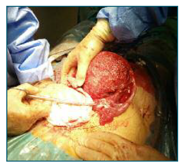
A primigravida in her early 30s, previously healthy, with an uneventful pregnancy except for the diagnosis of posterior placenta previa, for which she had no known risk factors, was admitted to the Emergency Department at 36 weeks and 1 day due to severe abdominal pain and regular uterine contractions. Gentle vaginal evaluation showed no vaginal bleeding or cervical dilation. Obstetric ultrasound showed a foetus in cephalic presentation with normal amniotic fluid and normal umbilical artery doppler velocimetry and a posterior placenta previa with no evidence of abruption. Cardiotocography showed no signs of foetal distress but regular uterine contractions. Persistence of intense complaints despite analgesic medication increased the suspicion of early-stage preterm labour. Due to the subsequent risk of placental haemorrhage, an immediate c-section was decided. When the abdominal cavity was reached and before the uterine incision, moderate volume hemoperitoneum was observed, mostly limited to the pelvic cavity and lower abdomen. The foetus was immediately extracted, with an Apgar Score of 9, 10 at the 1st and 5th minutes, respectively, and a weigh of 2790 g. The placenta was easily removed, showing no signs of previous detachment. Following the uterine incision closure, further abdominal cavity inspection revealed the only additional haemorrhagic source as an extensive lesion of friable and superficially vascularized tissue covering the posterior uterine wall, both adnexal regions and anterior wall of the rectum (Figure 1). Due to the macroscopic appearance and typical location, in spite of the extensive distribution, deciduosis was suspected, for which no biopsy was performed. Haemostasis was achieved with electrocoagulation and a reabsorbable oxidized cellulose meshwork. A drain was placed on the Douglas Pouch for blood loss surveillance. The postoperative hemoglobulin value was 9,2 g/dl (12 g/dl preoperative) and the patient was managed conservatively with iron supplementation. After an initial difficult pain management and constipation, she had a favourable evolution and was discharged on the 9th postoperative day wi-thout further complications. Puerperal evaluation at 4 weeks postpartum had no relevant findings, namely no lesions visualized on gynaecological ultrasound or fur-ther episodes of abdominal pain suggestive of intraabdominal bleeding. She recovered completely and had a second pregnancy 3 years later, also complicated by placenta previa. During the second c-section no deciduosis was described, but extensive intraabdominal adhesions were found.

Figure 1 Friable and highly haemorrhagic lesion covering the posterior surface of the uterus and both adnexal regions. 

Discussion

Ectopic decidualization is mostly an incidental finding during surgery, but may be the cause of severe hemoperitoneum and other haemorrhagic complications. The differential diagnosis of these lesions is malignancy (such as peritoneal mesothelioma) or tuberculosis. Due to the macroscopic resemblance of these diseases in some cases, biopsy should be done to stablish the diagnosis18. Whenever suspicion of this benign entity and surgical risk of biopsy are high, conservative haemostasis should be employed and unnecessary manipulation avoided due to the good prognosis and spontaneous regression of the lesions within four to six weeks postpartum5,6.

Authors’ contribution

DM and FS were directly responsible for the initial approach to the patient on the emergency department and surgical approach; DM, ISG, ML were closely involved in the follow up of the patient. All authors were major contributors in writing the manuscript. All authors read and approved the final manuscript.

Conflicts of interest

The authors have no conflicts of interest to disclose.

Informed consent

Patient consent was obtained in written.

References

1. Markou GA, Goubin-Versini I, Carbunaru OM, Karatzios C, Muray JM, Fysekidis M. Macroscopic deciduosis in pregnancy is finally a common entity. Eur J Obstet Gynecol Reprod Biol 2016 Feb;197:54-8 [ Links ]

2. Abramowicz S, Kouteich K, Grémain J, Sabourin JC, Marpeau L, Sergent F. Giant ectopic peritoneal and omental deciduosis mimicking a peritoneal carcinomatosis. Gynecol Obstet Fertil 2014 Mar;42(3):182-4 [ Links ]

3. Ng SW, Norwitz GA, Pavlicev M, Tilburgs T, Simón C, Norwitz ER. Endometrial Decidualization: The Primary Driver of Pregnancy Health. Int J Mol Sci 2020 Jun;21(11):4092. [ Links ]

4. Lepage F, Schramm B. Deciduosis of the uterine cervix. Gynecol Obstet 1955;54:550-63. [ Links ]

5. Semenova T, Metello J, Almeida M, Leite C, Santos A. Ectopic Decidualization as a cause of intra-caesarean hemorrhage. Acta Obstetrica e Ginecologica Portuguesa 2013;7(4):316-319 [ Links ]

6. Mendes J, Costa A. Ectopic Decidualization: A Forgotten Entity. Acta Med Port 2016 Jan;29(1):63-72 [ Links ]

7. Casanova J, Jurgiel J, Henriques V. Peritoneal deciduosis mimicking peritoneal Carcinomatosis: A case report. Gynecol Oncol Rep 2021 Jul 2;37:100827 [ Links ]

8. Masouridou S, Mamopoulos A, Mavromatidis G, Karagiannis V. Endometriosis and perinatal outcome - a systematic review of the literature. Curr Women's Health Rev 2012;8:121-30 [ Links ]

9. Buttner A, Bassler R, Theele C. Pregnancy-associated ectopic decidua (deciduosis) of the greater omentum. An analysis of 60 biopsies with cases of fibrosing deciduosis and leiomyomatosis peritonealis disseminata. Pathol Res Pract 1993 Apr;189(3):352-9 [ Links ]

10. Salehgargari S, Sahebdel B, Zare A, Abolhassani H. Ectopic Decidual Reaction Mimicking Irritable Bowel Syndrome: A Case Report. Acta Med Iran 2014;52(1):88-90 [ Links ]

11. Kondoh E, Shimizu M, Kakui K, Mikami Y, Tatsumi K, Konishi I. Deciduosis can cause remarkable leukocytosis and obscure abdominal pain. J Obstet Gynaecol Res 2012;38:1376-8. [ Links ]

12. Nishikawa A, Kondoh E, Hamanishi J, Yamaguchi K, Ueda A, Sato Y, Konishi I. Ileal perforation and massive intestinal hemorrhage from endometriosis in pregnancy: case report and literature review. Eur J Obstet Gynecol Reprod Biol 2013;170:20-4. [ Links ]

13. Aziz U, Kulkarni A, Lazic D, Cullimore JE. Spontaneous rupture of the uterine vessels in pregnancy. Obstet Gynecol 2004;103:1089-91. [ Links ]

14. Mizumoto Y, Furuya K, Kikuchi Y, Aida S, Hyakutake K, Tamai S, Nagata I. Spontaneous rupture of the uterine vessels in a pregnancy complicated by endometriosis. Acta Obstet Gynecol Scand 1996;75:860-2. [ Links ]

15. O'Leary SM. Ectopic decidualization causing massive postpartum intraperitoneal hemorrhage. Obstet Gynecol 2006;108:776-9. [ Links ]

16. Zaytsev P, Taxy JB. Pregnancy associated ectopic decidua. Am J Surg Pathol 1987;11:526-30. [ Links ]

17. Bolat F, Canpolat T, Tarim E. Pregnancy-related peritoneal ectopic decidua (deciduosis): morphological and clinical evaluation. Turk Patoloji Derg 2012;28(1):56-60. [ Links ]

18. Canlorbe G, Goubin-Versini I, Azria E, Abdul-Razak R, Muray JM. La déciduose ectopique: variabilités de présentation pendant la grossesse et diagnostics différentiels [Ectopic decidua: variability of presentation in pregnancy and differential diagnoses]. Gynecol Obstet Fertil 2012 Apr;40(4):235-40. [ Links ]

Received: April 26, 2022; Accepted: May 28, 2022

Correspondence to: Diana Margarida Monteiro E-mail: diana_monteiro_@hotmail.com

Creative Commons License This is an open-access article distributed under the terms of the Creative Commons Attribution License