


 Inglês (pdf)
 Artigo em XML
 Referências do artigo
 Enviar este artigo por email
 Citado por SciELO
  Similares em
SciELO
Similares em
SciELO 
 Permalink
Introduction
Preeclampsia is a pregnancy-associated disorder classically defined as newly-onset hypertension and proteinuria after 20 weeks’ gestation and most frequently in the third trimester1. The onset of hemolysis, elevated liver enzymes and low platelet count (HELLP) Syndrome is one of the more severe and rarer forms of preeclampsia and is associated with even higher rates of maternal morbimortality1-4. When symptoms associated with hypertensive disorders of pregnancy (HDP) arise before 20 weeks’ gestation, the presence of a molar pregnancy should be excluded.
Molar pregnancy or hydatidiform mole (HM) is a premalignant disease that arises from gestational tissue and is classified as either partial (PM) or complete mole (CM)5. A PM is formed by triploid cells that most commonly result from the fertilization of an ovum by two sperms or, more rarely, a diploid sperm5,6. In this entity, foetal tissue is present and cardiac activity may develop.
Ultrasound diagnosis is challenging, with the majority of cases remaining unsuspected after routine ultrasound examination, where a foetus with vitality and regular size may be visualized and placental hydropic structures or increased echogenicity absent or subtle7-9. These changes may become more evident in the second trimester, which may include a mildly growth-restricted foetus with proportionate head size or slight microcephaly and an enlarged and multicystic placenta10. Therefore, only 18-29% of cases are diagnosed before histopathological evaluation, being misdiagnosed as spontaneous or incomplete abortion8,11,12. PM displays elevated β-human chorionic gonadotropin (β-hCG), but is significantly less noticeable than CM and therefore less likely to present with hyperemesis, hyperthyroidism or theca lutein cysts11.
Despite the high frequency of early miscarriage due to the underlying chromosomal anomaly, some pregnancies continue to progress13. It is uncommon for a PM to reach the second trimester, but in such cases of a live foetus the risk of preeclampsia is increased, therefore diagnosis should be made as soon as possible to prevent the severe risk of maternal morbimortality11. Should the diagnosis of PM be made with a live foetus, taking into account the serious threat to the mother’s health, couples should be advised to terminate the pregnancy without delay after maternal stabilization4,6,11,14.
Case Report
We present the case of a primigravida in her early 30s, with previously known well-controlled asthma and no other relevant medical history. At 17 weeks gestation, she was transferred to our Hospital from her Primary Health Care Unit where severe hypertension (220/180 mmHg) was detected on a routine visit. She complained of peripheral oedema up to the knees, with progressive worsening, over the last 2 weeks. Additionally, she reported episodes of epigastric pain, indigestion and vomiting during the same period. She denied having headaches, blurred vision or other accompanying symptoms. On physical examination, she presented persistent severe hypertension (185/120 mmHg) and bilateral soft peri-malleolar oedema. She had no other relevant findings, namely neurologic symptoms, ocular changes, or genital bleeding (Figure 1).

On the ultrasound, the foetus had good vitality and normal amniotic fluid and foetal biometry but agenesis of the corpus callosum and bilateral ventriculomegaly was suspected (Figure 2). The placenta was enlarged and showed numerous cystic lesions (Figure 3). On the transvaginal ultrasound, both ovaries were enlarged (56- and 64-mm largest diameter, respectively), with multiple cysts, suggestive of theca lutein cysts (Figure 4). The urine dipstick test showed 3+ for proteinuria. Blood analysis showed hemoglobin 11.8 g/dl, platelet count was 163 (150-400) 109/L, aspartate aminotransferase activity (AST) 179 (<34) U/L, alanine aminotransferase activity (ALT) 114 (<55) U/L), lactate dehydrogenase (LDH) 454 (<220) U/L and uric acid 6.5 (<6.0) mg/dL, thyroid stimulating hormone (TSH) 0.23 (0.35-4.94) µUI/mL, but normal free thyroxine (T4) and severely increased β-hCG (646.000 mUI/ml), with no other relevant changes.
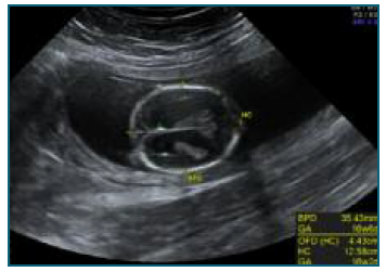
Figure 2 Transventricular view of foetal cranium at 17 weeks gestation - suspected agenesis of corpus callosum and bilateral ventriculomegaly.
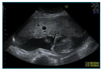
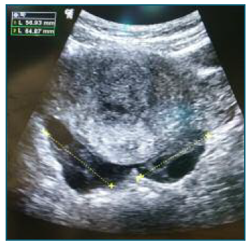
Figure 4 Transabdominal view after uterine evacuation - bilaterally enlarged ovaries (56- and 64-mm largest diameter, respectively), with multiple cysts, suggestive of theca lutein cysts.
Upon review of prenatal records undertaken in another hospital, the first-trimester ultrasound at 12 weeks and 1 day showed no relevant morphologic foetal or placental anomaly and had nuchal translucency (NT) below the 95th percentile. The combined screening for foetal aneuploidy showed an increased risk for trisomy 21 (1:157) due to increased β-hCG (7,39 multiples-of-median (MoM)) and after counselling at the time, the couple had chosen to perform a non-invasive prenatal test (cell-free foetal DNA (cfDNA)) instead of an amniocentesis, which was negative for trisomy 21, 18 and 13.
The combination of symptoms (hypertension and peripheral oedema), analytical abnormalities (elevated β-hCG), typical cystic lesions of the placenta and theca-lutein ovarian cysts rapidly pointed the team to the diagnosis of a probable HDP in a PM.
She was admitted to the intensive care unit due to the difficult management of hypertension. She was medicated with labetalol protocol, with suboptimal effect and less than 12 hours later her general medical condition worsened, with anemia (8.9 g/dl hemoglobin), thrombocytopenia 64×109/L, AST/ALT 612/285 U/L, LDH 1265 U/L and slightly elevated bilirubin. After multidisciplinary counselling of the couple, termination of the pregnancy was decided due to findings consistent with HELLP syndrome in a pregnancy before the limit of foetal viability. An ultrasound-guided dilation and evacuation under general anesthesia were performed without complications. Magnesium sulfate perfusion was initiated for 24 hours and hypertension was managed with labetalol and posteriorly captopril and amlodipine.
After pregnancy evacuation and initial worsening of anemia (nadir 6.2 g/dl hemoglobin) and thrombocytopenia (nadir 30×109/L), requiring transfusion of one unit of red blood cells, she progressively improved all clinical parameters and was discharged after 8 days. Foetal necropsy showed arachnodactyly of the hands, bilateral “sandal gap” and camptodactyly of the 2nd and 4th toes of the left foot and foetal karyotype revealed a 69, XXY. β-hCG became negative after 2 months and she maintained analytic surveillance for another 6 months. Four years later, she had a healthy baby, with no pregnancy complications.
Discussion
HDP are a major cause of maternal and foetal morbimortality and every effort should be made to screen, treat and manage these women with the utmost care1. New-onset hypertension or worsening of chronic hypertension during pregnancy, before 20 weeks gestation, should prompt exclusion of maternal underlying conditions that may worsen with pregnancy, such as antiphospholipid syndrome, thrombotic thrombocytopenic purpura, hemolytic-uremic syndrome, systemic lupus erythematosus and other autoimmune diseases and renal conditions1,15. With the introduction of ultrasound as part of routine evaluation of pregnant women, the frequency of late diagnosis of HM and its associated complications have decreased. Nonetheless, preeclampsia-like symptoms in a pregnant woman before 20 weeks mandates exclusion of molar pregnancy8. In our case, the patient had a PM that progressed into the second trimester and eventually developed one of the most severe forms of the HDP spectrum - a HELLP Syndrome. It is a rare condition that poses an even higher risk of morbimortality than other HDP.
Early diagnosis of PM and CM does not result in reduction of the risk of progression to gestational trophoblastic neoplasia, but early evacuation significantly decreases the risk of clinical complications such as preeclampsia, so every effort should be made to diagnose these cases as early as possible12,20.
This couple chose to perform a cfDNA test, due to the lower risk of abortion compared to an invasive technique, which was negative for the specific chromosomal abnormalities screened (trisomy 21, 18 and 13). Instead, the performance of an invasive technique would have resulted in an earlier diagnosis of triploidy on the karyotype.
The debate has emerged over the role of cfDNA as the primary screening program for aneuploidies and the potentially missed foetal anomalies beyond trisomy 21, 18 and 13 versus combined first-trimester screening based on ultrasound evaluation (NT and foetal heart rate (FHR)) and maternal serum biochemical markers (free β-hCG and pregnancy-associated plasma protein-A (PAPP-A)) 16,17.
Regarding sonographic markers (NT or FHR), triploid foetuses do not display significant differences to euploid foetuses18. As for biochemical markers, though free β-hCG may be elevated in trisomy 21, leading to a high-risk result in the first-trimester screening test for aneuploidies, severe elevations should raise suspicion of other anomalies, such as diandric triploidy (paternal origin) 12. According to the literature, free β-hCG on trisomy 21 foetuses deviates from euploid 2.2 MoM (range 1.5-3.3 MoM), where diandric triploid foetuses deviate 8.7 MoM (range 2.8-47.0 MoM) 10,17.
Analysis of the first-trimester screening result should go beyond the risk value for trisomy 21,18 and 13. Careful ponderation of free β-hCG and PAPP-A values should be independently interpreted and contextualized. 17,19
This clinical presentation of a serious condition, such as the HELLP Syndrome, on a currently rare diagnosis on the 2nd trimester, highlights the importance of being aware of the possibility of a PM, in order to advise rapid termination of pregnancy, given its high risk of morbimortality. In addition, this case makes us ponder the role of each obstetric innovation and the importance of an adequate counselling of cfDNA limitations.
With each generation, Fetal Medicine and Prenatal Diagnosis Specialists have more knowledge and diagnostic tools at their disposal. However, these innovations require clinical sense and wisdom for a correct interpretation. Prenatal counselling of the couple should be done based on all parameters to advise for either routine surveillance, expectant management, cfDNA test or invasive technique (either chorionic villus sampling or amniocentesis).
Author contribution statement
DM, MD and ISG were directly responsible for the initial approach to the patient in the emergency department and surgical approach; JPN was responsible for the long-term follow-up of the patient. All authors were major contributors to writing the manuscript. All authors read and approved the final manuscript.

