SciELO - Scientific Electronic Library Online

 
vol.17 número2Migração de dispositivo intrauterino em posição invertida índice de autoresíndice de materiabúsqueda de artículos
Home Pagelista alfabética de revistas  

Servicios Personalizados

Revista

Articulo

Indicadores

Links relacionados

  • No hay articulos similaresSimilares en SciELO

Compartir


Acta Obstétrica e Ginecológica Portuguesa

versión impresa ISSN 1646-5830

Acta Obstet Ginecol Port vol.17 no.2 Algés jun. 2023  Epub 30-Jun-2023


Issue Image/Imagem do Trimestre

Not everything is always genetic - A prenatal presentation of tortuous and aneurysmal ductus arteriosus

Nem tudo é sempre genético - Apresentação pré-natal de canal arterial tortuoso e aneurismático

Raquel Gouveia Silva1 
http://orcid.org/0000-0002-5470-5490

Márcia Rodrigues1 

Mónica Rebelo2 

1. Serviço de Genética Médica, Departamento de Pediatria, Hospital de Santa Maria, Centro Hospitalar Universitário Lisboa Norte, Lisboa, Portugal.

2. Serviço de Cardiologia Pediátrica, Departamento de Pediatria, Hospital de Santa Maria, Centro Hospitalar Universitário Lisboa Norte, Lisboa, Portugal.


Abstract

Tortuous and aneurysmal ductus arteriosus (TADA) is a relatively rare condition, especially in the first and second trimesters. The ductus arteriosus has functional importance in the fetal circulation, being its aneurysmatic format characterized by saccular dilatation of the vessel. Majority of cases have a benign evolution, although they can be associated with syndromic forms and with important complications. We report the clinical management and echocardiographic follow-up in a newborn with this very early finding in prenatal setting.

Keywords: Aneurysmal ductus arteriosus; Prenatal setting; Early diagnosis

Resumo

O canal arterial tortuoso e aneurismático (CATA) é uma condição relativamente rara, especialmente no primeiro e segundo trimestres. O canal arterial tem importância funcional na circulação fetal, sendo seu formato aneurismático caracterizado pela dilatação sacular do vaso. A maioria dos casos tem evolução benigna, embora possam estar associados a formas sindromáticas e a complicações importantes. Relatamos a orientação clínica e o seguimento ecocardiográfico num recém-nascido com este achado muito precoce em contexto pré-natal.

Palavras-chave: Canal arterial aneurismático; Contexto pré-natal; Diagnóstico precoce

Case

Prenatal diagnosis of TADA is usually incidental in third trimester. The vast majority of cases has a good prognosis, though regular ultrasound follow-up is needed given the associated thrombotic risk1. Maternal diabetes and blood group A are considered high-risk factors1,2. TADA is related with trisomy 21 and 13, as well as, connective tissue diseases, as Marfan and Ehlers-Danlos syndromes3, related with alterations in elastin expression or intima cushioning, or has multifactorial etiology1,2.

We present a male with prenatal diagnosis of TADA. His mother, a 36-year-old woman, with blood group A, has a personal history of hypothyroidism and type 1 diabetes with no history of exposure to teratogens, namely consumption of polyphenol-rich substances, herbal infusions, or nonsteroidal anti-inflammatory drugs4, and has a previous healthy daughter.

At 12 weeks of gestation, fetal ultrasound revealed a small omphalocele with intestinal content and invasive prenatal testing was performed through chorionic villus sampling. The results of QF-PCR and arrayCGH (CGX-HD 180K, PerkinElmer) were both normal. MS-MLPA for Beckwith-Wiedemann syndrome was not possible to perform due to insufficient fetal DNA and early gestational age. Subsequent ultrasound evaluation showed regression of the omphalocele and adequate fetal growth.

Considering maternal diabetes background and the need to exclude other congenital malformations, fetal echocardiogram was performed at 24 weeks. It showed a tortuous ductus arteriosus with a turbulent high velocity flow, with a maximum velocity of 130 cm/s (Figure 1). Regarding the potential thrombotic or rupture risks, weekly echocardiogram was performed until birth, evaluating the ductus morphology and doppler flow, and searching for signs of right ventricular disfunction (tricuspid regurgitation and ventricular dilatation), showing no deterioration. The child was born at 38 weeks by vaginal delivery, with a birth weight of 3150 grams (25th centile) and Apgar score 9/10. Echocardiogram performed on the 3rd day of life, corroborated the prenatal findings, with complete resolution at 1 month of age (Figure 2).
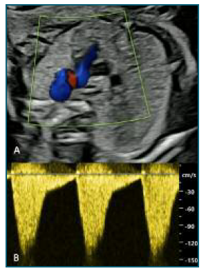
Figure 1 Prenatal ultrasound evaluation, twenty-four weeks gestation prenatal transabdominal echo: A - Three vessel view showing tortuous and aneurismatic ductus arteriosus; B - Flow Doppler with elevated velocity (120-130 cm/s). 
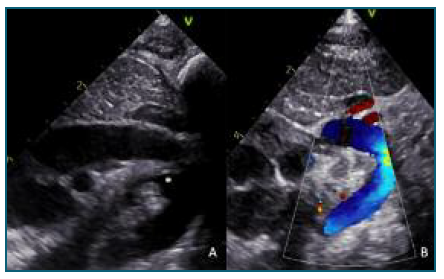
Figure 2 Postnatal ultrasound evaluation (Suprasternal views) A - Three days of life postnatal transthoracic echo: aortic arch with ductus arteriosus (large ampola*) and restriction at pulmonar side; B - One month of life postnatal transthoracic echo: normal aortic arch, no ductus arteriosus (closed). 

This case highlights the unusual frequency of TADA in the earlier stages of pregnancy and recalls its most frequently observed follow-up and prognosis.

Author contributions

All authors made a substantial contribution to the information or material submitted for publication.

Declarations of interest

The authors have no conflicts of interest to declare.

Informed consent

Patient consent for publication was obtained.

References

1. Sequeira AT, Lemos M, Palma MJ. Prenatal thrombosis of the ductus arteriosus. Cardiol Young. 2019 Mar;29(3):408-409. DOI: https://doi.org/10.1017/S1047951118002159. Epub 2018 Dec 26. [ Links ]

2. Pereira AG, Teixeira A, Martins FM. Spontaneous thrombosis of the ductus arteriosus. Rev Port Cardiol 2011; 30: 537-540. [ Links ]

3. Ganesan S, Hutchinson DP, Sampson AJ. Prenatal diagnosis of ductus arteriosus aneurysm. Ultrasound. 2015 Nov;23(4):251-3. DOI: https://doi.org/10.1177/1742271X15587931. Epub 2015 May 27. PMID: 27433265; PMCID: PMC4760603. [ Links ]

4. Paulo Zielinsky, Antonio Luiz Piccoli, João Luiz Langer Manica & Luiz Henrique Soares Nicoloso (2010) New insights on fetal ductal constriction: role of maternal ingestion of polyphenol-rich foods, Expert Review of Cardiovascular Therapy, 8:2, 291-298, DOI: https://doi.org/10.1586/erc.09.174 [ Links ]

Received: July 30, 2022; Accepted: October 27, 2022

Correspondence to: Raquel Gouveia Silva E-mail: anaraquelsilva91@gmail.com

Creative Commons License This is an open-access article distributed under the terms of the Creative Commons Attribution License