


 Inglés (pdf)
 Articulo en XML
 Referencias del artículo
 Enviar articulo por email
 Citado por SciELO
  Similares en
SciELO
Similares en
SciELO 
 Permalink
Introduction
Clinical hyperthyroidism occurs in 0.2-0.7% of pregnancies, and Graves’ disease (GD) is responsible for about 95% of these cases1,2.
Due to both thyrotropin receptor antibodies (TRAb) and antithyroid drugs (ATDs) crossing the placenta, treatment of Graves’ disease can be a challenge3. It is necessary to balance control of maternal hyperthyroidism while maintaining a euthyroid state in the fetus3. In some cases, the maternal overtreatment with ATDs may be responsible for fetal hypothyroidism, which can be suspected by the enlargement of the fetal thyroid gland on prenatal ultrasound2,4.
The authors describe a case of prenatal diagnosis and management of fetal goiter related to maternal Graves’ disease.
An informed consent was obtained from the patient before writing this manuscript.
Case report
A 26-years-old caucasian pregnant woman, G2P1 (previous gestation without complications) was referred to a tertiary center due to Graves’ disease, at 12 weeks gestation. The patient was diagnosed after her first pregnancy and was treated with methimazole since then. At 12 weeks, her thyroid function was not satisfactorily controlled. TRAb level was 24 U/L (normal range: < 1U/L), so the treatment with a higher dose of methimazole was necessary. At 25 weeks, due to a normalization of her thyroid function, the necessary dose of methimazole was lower.
Fetal ultrasound scans at 12, 21 and 29 weeks of gestation were normal. The 32 week ultrasound identified an enlargement of fetal thyroid gland, both diameter and circumference superior to the 95th percentile (Figure 1), and hyperextension of the fetal neck Figure 2). No other findings were seen, to help the differential diagnosis between fetal hypo or hyperthyroidism. As the fetal thyroid enlargement persisted, cordocentesis was performed at the 34 weeks to determine fetal thyroid status, which revealed fetal hypothyroidism [thyroid stimulating hormone (TSH) was 75 uUI/mL (normal range: 0,4 to 4 uUI/mL) and free thyroxine (FT4) was 0,62 ng/dL (normal range: 0,7 to 1,5 ng/dL)]. Maternal therapy with methimazole was suspended and 0,4 mg of levothyroxine was administered into the amniotic fluid. After one week, fetal thyroid presented a decreasing of its biometry (28% in thyroid circumference), and fetal thyroid function improved (TSH of 0,53 uUI/mL). Fetal therapy was performed with weekly intra-amniotic injections of 0,4 mg of levothyroxine until 36 weeks, and amniotic fluid levels of TSH and FT4 were obtained each time.
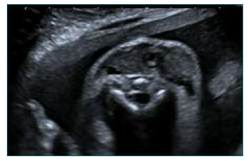
Figure 1 Fetal goiter: ultrasound transverse view of the fetal neck at 32 weeks, showing an anterior solid homogeneous bilobed mass, that is symmetric around the trachea.
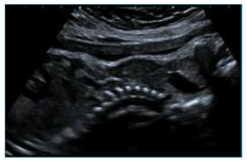
Figure 2 Fetal goiter: ultrasound sagital view of the fetal neck at 32 weeks, showing an enlargement of the thyroid causing hyperextension of the neck.
At 37 weeks and 4 days, the patient was admitted in spontaneous labor, resulting in vaginal delivery. The male newborn weighted 2385 gr and the Apgar score was 8/8/10. Umbilical cord blood sample showed: TSH of 10 UI/mL (normal range: 0.70 to 15.20 UI/mL), FT4 of 0,75 ng/dL (normal range: 1.05 to 3.21 ng/dL) and TRAb of 13 U/L (normal range: < 0.1 U/L). He was discharged at day 4. On day 15 after delivery, the newborn had normal thyroid function and the TRAb level was 9.9 IU/L. At 1 year-old, the infant has normal growth and no evidence of psychomotor development delay.
Discussion
Maternal Graves’ disease is the most common cause of fetal goiter and, if not properly managed, can result in severe maternal, fetal, and neonatal morbidity and mortality5.
Fetal goiter is defined as an abnormal enlargement of the fetal thyroid gland circumference or diameter above the 95th percentile) 6. It is found in about 19% of the fetuses carried by mothers with GD, and it indicates thyroid dysfunction6. However, fetal goiter can be present not only in cases of fetal hyperthyroidism, but also in fetal hypothyroidism3,6. Other ultrasound findings that can be present in cases of fetal hyperthyroidism are fetal tachycardia, growth restriction and nonimmune fetal hydrops3,4. In extreme cases, hearth failure can cause fetal death3.
Ultrasound combined with color Doppler is helpful for the initial diagnosis and monitoring, but has limited ability to discriminate between fetal hyperthyroidism and hypothyroidism5. Cordocentesis is the gold standard method for confirming fetal thyroid hormone levels for a fetal thyroid diagnosis6.
This case showed that, the balance between the risk of fetal hypothyroidism due to ATD placental passage and fetal hyperthyroidism due to TRAB placental passage, can be difficult to manage in Graves’ disease.
Fetal hypothyroidism should be thoroughly evaluated, diagnosed and treated, as it is a treatable cause of delayed psychomotor development. Fetal therapy can be done not only by administering drugs to the mother, but also by injecting levothyroxine into the amniotic fluid, which does not cross the placenta4,6.

