


 Inglés (pdf)
 Articulo en XML
 Referencias del artículo
 Enviar articulo por email
 Citado por SciELO
  Similares en
SciELO
Similares en
SciELO 
 Permalink
Congenital diaphragmatic hernia (CDH) is one of the most severe birth defects, with extremely high neonatal mortality and morbidity. It has a prevalence of 1/3000 live births. In about 75 to 90% of cases the defect has a left-sided predominance (10 - 15% to the right; 1-2% bilateral)1.
CDH consists of a congenital defect of the dia-phragm that results in pulmonary hypoplasia due to herniation of intra-abdominal viscera into the thorax. The absence of diaphragmatic continuity at the 10th week of pregnancy is always related to the abnormal position of adjacent organs: the intestines are usually involved; in left-sided hernia, the stomach is often present, while the liver is more often herniated when the defect is on the right side. Abnormal development of the lung starts at 14-16 weeks, the diminished proliferative capacity in CDH results in lung hypoplasia, pulmonary hypertension and a dysfunctional surfactant system1.
Antenatal ultrasound diagnosis its feasible in 60% of the cases, with a mean gestational age of 22-24 weeks thus allowing an attempted in utero referral to a tertiary care center2.
In this case the ultrasonography at 23 weeks was normal (Figure 1). Surprisingly, in the third trimester ultrasound performed at 32 weeks, it was possible to unequivocally observe the presence of the stomach and intestine in the fetal four chambers view, as well as deviation of the cardiac axis to the left, raising the suspicion of left -sided CDH (Figure 2). This was a second pregnancy of a healthy couple with no relevant family history and a healthy first-born child.
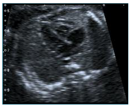
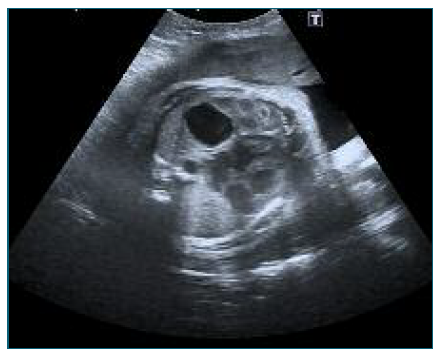
Figure 2 Axial plane view of the fetal thorax at 32 weeks: stomach and bowel were clearly visible at the level of the “4-chamber view” with a right heart deviation, making the diagnosis of left-sided congenital diaphragmatic hernia.
The most reliable method of lung estimation and marker of postnatal morbidity and mortality is o/e LHR. In our case, the o/e LHR was calculated on the 4-chamber view using a web-based calculator (www.totaltrial.eu) with the anteroposterior diameter of the lung. 1) The o/e LHR was 32 % and the estimated postnatal survival rate was 40-60%2. The absence of other structural abnormalities and herniation of the liver alone also contributed to a favorable prognosis. Genetic study was carried out postnatally due to gestational age and because it would not change the clinical conduct, given the good prognosis of the case. The couple was referred to a tertiary care center, where spontaneously delivered at 37 weeks. The surgery was performed 2 days later with good results. The baby has presently 3 months with a normal development and parents decided not do postnatal genetic study.
The etiology of CDH is still unknown1. Like in this case, this structural defect is often sporadic, without a family history. Management of CDH remains challenging: the prognosis could be improved by fetal endoscopic tracheal occlusion (FETO) an experimental technique which corrects the major problems of this condition - pulmonary hypoplasia and hypertension. In Portugal, postnatal surgical repair after neonate stability is the only option3.
Ultrasound is crucial for prenatal diagnosis, prediction of disease outcomes and to the program management and delivery. This case demonstrates that this pa-thology can, in rare cases, only manifest itself late in pregnancy and how important is a systematic morphological study the third trimester - timely diagnosis and referral to a third level hospital is crucial to the survival of newborns.

