


 Inglês (pdf)
 Artigo em XML
 Referências do artigo
 Enviar este artigo por email
 Citado por SciELO
  Similares em
SciELO
Similares em
SciELO 
 Permalink
Introduction
Vasculitides are characterized by a varied group of diseases capable of triggering inflammation in the walls of blood vessels with or without necrosis.1 These conditions are usually caused by the deposition of immune complexes in the endothelium, which activates the complement system responsible for stimulating mast cell degranulation and neutrophil chemotaxis, culminating in vascular wall damage.2 Leukocytoclastic vasculitis (LV) is a small vessel vasculitis that mainly affects venules, characterized by deposition of immune complexes, fragmentation of neutrophil nuclei and inflammatory infiltrate with resultant fibrinoid necrosis.3,4 LV affects equally both genders and can affect patients of all ages.4
The present work reports a case of LV that manifests itself mainly through cutaneous ulcers of difficult remission, approaching the clinic of the observed phenomenon and the disease. This study met all the criteria recommended by resolution 166/2018 - CONEP, Brazil. It was properly evaluated and approved by the Research Ethics Committee under CAAE 54238221.2.0000.5219 and Consolidated Opinion 5.306.012.
Case report
A 54-year-old female patient reported, in August 2019, severe pain and swelling in lower limbs, mainly below knees and left leg, associated with intense pruritus and local paresthesia. Patient’s past medical history included: obesity and previous surgical procedure of total thyroidectomy due to a suspicious nodule in the thyroid 5 years ago, currently using levothyroxine.
On physical examination, we observed large veins in both lower limbs (Clinical, Etiology, Anatomy and Pathophysiology (CEAP) C2 on right and C3 on left lower limb). In addition, there were wounds at the dorsum of the feet and medial malleolus. Pulses were bilaterally preserved.
Patient brought a biopsy result of the erythematous-purpuric lesions in the lower limbs requested by a rheumatologist in 2018, with the diagnosis of acute leukocytoclastic vasculitis. Patient reported that, in that time, corticosteroid was prescribed, but she did not remember the dosage prescribed. She also reported that she discontinued the treatment by her own initiative a short time later.
Diagnostic analytic tests were requested to evaluate the condition, including Hemogram, Erythrocyte Sedimentation Rate and C-Reactive Protein Chain, with normal results. Antinuclear Factor presented a result of 1:80 (with a positive laboratory result for >1:80). A Rheumatologist consultation was requested and Systemic Lupus Erythematosus (SLE) diagnosis was ruled-out.
The initial treatment included phlebotonics, moisturizing lotions, prednisone 20 mg/day, paracetamol with codeine and compressive stockings.
The patient returned for an appointment in December 2020, reporting that she had not performed the prescribed treatment and referring aggravation of the ulcers and pain in the lower limbs. On physical examination, erythematous-purpuric macular lesions were observed on the legs, ankles and feet, in addition to multiple irregular ulcerations with a secretive and necrotic background at the dorsum of the feet and medial malleolus (Figure 1). Doppler ultrasound showed segmental reflux at the right saphenofemoral joint, also affecting the right anterior accessory saphenous vein and significant reflux at the left great saphenous vein.
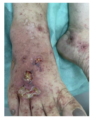
Figure 1 Presence of ulcers on the dorsum of the feet and medial malleolus, in addition to erythematous-purpuric macular lesions.
Treatment implemented by the Rheumatology department included vasodilators, increasing prednisone dosage to 40 mg/day, methotrexate 10 mg/week, azathioprine 50 mg/day, folic acid, analgesics and bandages with chlorhexidine acetate to treat the ulcers. Antistreptolysin O (ASO) titer confirmed group A streptococcal infection and levofloxacin was prescribed for 10 days.
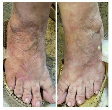
After four months, the patient returned to an appointment with a good evolution of the ulcers (Figure 2) and significant improvement of the pain. The medications prescribed before were maintained and an antihistamine medication was associated for pruritus.
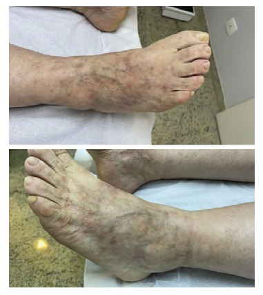
In July 2021, the patient returned to the clinic presenting complete healing of the ulcerative and macular lesions (Figure 3) and remission of pain and pruritus. She was instructed about personal care, vasodilators were maintained and the dosage of corticosteroids was gradually withdrawn to 5 mg/day to avoid recurrence of the wounds.
Finally, at the end of 2021, the patient was referred to a vascular surgeon and underwent a radiofrequency thermoablation of saphenous veins associated with phlebectomy of tributary veins, leading to improvement of the swelling and local paresthesia.
Discussion
Despite of LV being an idiopathic condition in most cases, it is necessary to exclude secondary causes, such as use of medication, infections, sepsis and malignancy.4 Therefore, disease diagnosis aims to determine the type of vasculitis and, when possible, its underlying cause.5 Besides that, the management of ulcerated lesions associated with LV is multidisciplinary and involves the orchestrated action of different medical specialties, such as Pathology, Rheumatology and Angiology, in order to diagnose and control the underlying disease and to treat and prevent wounds recurrence.
With the purpose of identifying characteristics of LV, triggering factors of the disease and excluding differential diagnoses, a detailed anamnesis and physical examination are crucial.2,6 Clinical suspicion should be confirmed through laboratory and/or instrumental tests, such as immunological investigation for vasculitis and connective tissue diseases, coagulation tests, lesional biopsy and histological tests.6,7 In the case described, direct immunofluorescence was not performed and could be useful in the classification of vasculitis. However, the presence of immunoglobulins is generally scarce in the lesions, making it questionable to use immunofluorescence as a routine test.3
Initially, conservative measures, such as bed rest, warming, elevation of the lower limbs and use of compressive stockings can reduce the deposition of immune complexes and help in wound healing.2,5,8 The use of topical corticosteroids and moisturizers can reduce pruritus and skin discomfort.8 Nonsteroidal anti-inflammatory drugs, analgesics and antihistamines can also be used to reduce pruritus and burning.2,8
Corticosteroids help in the resolution of painful ulcers and prevent the appearance of new lesions, however, long-term therapy is not recommended in chronic vasculitis due to the risk of complications with treatment and exacerbation of the lesions due to increased susceptibility to infections.8 Patients with vasculitis lasting more than 4 weeks or who respond incompletely to the use of corticosteroids can use corticosteroid sparing agents, such as colchicine and dapsone.5,8
Superinfection of open lesions is an imminent risk and the treatment of ulcers must be based on containing the contamination. Systemic antibacterials and topic agents, such as creams containing silver sulfadiazine or cadexomer-iodine ointment, can be used to treat the infection and remove deposited necrotic tissue. Bandages are not recommended in case of ulcer infection.8
In order to reduce the chance of relapse of ulcerative lesions, it is important to avoid possible triggering agents, such as infections and medications. In cases of recurrence of vasculitis and ulcers, prednisone can be used, reducing the dosage and gradually replacing it with more potent immunosuppressants, such as methotrexate or azathioprine. 9
Regarding the surgical treatment of chronic venous disease, it is indicated for patients with disabling and persistent discomfort in lower limbs or in cases of venous ulcers that are difficult to heal, as well as for cases of recurrence of varicose veins and patients with difficulty in adhering to treatment with compression elastic stockings. Surgery for incompetent truncal veins is indicated in order to avoid blood reflux overload on the superficial venous system. As for the tributary veins that have communication with the incompetent saphenous vein, it is possible to perform the avulsion of those concomitant with a saphenous vein ablation procedure.10
Thermal ablation using radiofrequency, performed in the case described, has efficacy and safety comparable to ligation and removal of incompetent veins. In addition to presenting low rates of complications and venous thromboembolism, radiofrequency ablation presents less technical failure than sclerotherapy with polidocanol and faster recovery, with less chance of ulcer recurrence, when compared to surgery.10
The case presented shows that the treatment of cutaneous ulcers associated with vasculitis can be long-lasting and requires a multidisciplinary approach and the use of different medications. Patient’s adherence to treatment is extremely important for a good prognosis, since non-compliance with treatment leads to relapse and worsening of pain and ulcers. Finally, radiofrequency thermoablation is shown to be a safe and effective option for treatment of incompetent saphenous veins, with less recurrence of cutaneous ulcers compared to surgery.

