SciELO - Scientific Electronic Library Online

 
vol.18 número4Lower extremity vasculitic ulcers - Case ReportGiant aneurysm originating from the costocervical trunk in a patient with aortic coarctation índice de autoresíndice de assuntosPesquisa de artigos
Home Pagelista alfabética de periódicos  

Serviços Personalizados

Journal

Artigo

Indicadores

Links relacionados

  • Não possue artigos similaresSimilares em SciELO

Compartilhar


Angiologia e Cirurgia Vascular

versão impressa ISSN 1646-706X

Angiol Cir Vasc vol.18 no.4 Lisboa dez. 2022  Epub 28-Fev-2023

https://doi.org/10.48750/acv.536 

Vascular Image

Autopsy image following aortic arch parallel grafting of a ruptured aortic aneurysm

Ryan Gouveia e Melo1 
http://orcid.org/0000-0001-8327-6384

Ruy Fernandes e Fernandes1 
http://orcid.org/0000-0003-1619-8851

Luís Silvestre1 

Luís Mendes Pedro1 
http://orcid.org/0000-0003-4310-9324

José Fernandes e Fernandes1 
http://orcid.org/0000-0002-2144-0430

1 Department of Vascular Surgery, Centro Hospitalar Universitário Lisboa Norte; Faculdade de Medicina da Universidade de Lisboa; Centro Cardiovascular da Universidade de Lisboa (CCUL@RISE). Portugal.


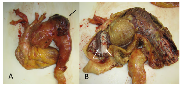
An 85-year-old man was admitted with a ruptured thoracic aortic aneurysm involving the mid/distal arch (bovine aortic arch) and proximal descending thoracic aorta. An emergent endovascular repair was performed with zone 0 TEVAR using a COOK® Zenith Thoracic Alpha endograft and endo-debranching of the innominate artery (IA) and left carotid artery (LCA) using parallel (chimney) grafts through right axillary and LCA cutdown access. An iliac limb (COOK® ZSLE) for the IA and a Viabahn (GORE®) for the LCA were used and both chimneys were relined using a self-expandable bare-metal stents. The left subclavian artery was covered and coiled. Although the rupture was sealed, the patient developed post-operative respiratory insufficiency, pneumonia and atelectasis related to the hemothorax. He died due to multiorgan failure at the 26th post-operative day. An autopsy was performed, where the aortic aneurysm with the ruptured hematoma is evident (A, arrow) and the position of the endograft, innominate artery chimney (B, arrow) and left carotid artery chimney (B, dotted arrow) can be seen in a ballerina position.

Fig A e B 

Received: February 04, 2023; Accepted: February 06, 2023

Corresponding Author: Ryan Gouveia e Melo, MD | fryan@campus.ul.pt Hospital de Santa Maria - Centro Hospitalar Universitário Lisboa Norte, EPE, Serviço de Angiologia e Cirurgia Vascular. Avenida Professor Egas Moniz, 1649-028, Lisboa, Portugal.

Creative Commons License This is an open-access article distributed under the terms of the Creative Commons Attribution License