


 Inglês (pdf)
 Artigo em XML
 Referências do artigo
 Enviar este artigo por email
 Citado por SciELO
  Similares em
SciELO
Similares em
SciELO 
 Permalink
Introduction
Dissection of main visceral arteries (celiac, superior mesenteric, and inferior mesenteric arteries) is usually a result of the extension of a dissection of the aorta to those arteries. However, spontaneous isolated visceral artery dissection is a quite rare condition and designates a dissection that originates in the visceral artery itself with no involvement of the aorta. We herein review the exceptional phenomenon of spontaneous isolated celiac and splenic artery dissection.
Methods
A non-systematic literature search was performed using the PubMed database. Only English literature was considered. Articles of interest were selected by the authors based on the title and abstract. Only publications reporting on isolated celiac artery dissection, isolated splenic artery dissection, or both, were considered. Further literature was obtained by cross-referencing. A narrative review was constructed, with the following structure: epidemiology; etiology; diagnosis; treatment; and follow-up.
Results
Epidemiology
Spontaneous isolated visceral artery dissections are very rare and must be distinguished from dissections resulting from the extension of a primary aortic dissection, which are much more common. Nevertheless, they are becoming more frequently diagnosed, probably as a result of a growing number of performed imaging techniques, particularly computed tomography angiography, with increasing quality.
The superior mesenteric artery is, due to unknown reasons, the most commonly involved visceral artery. Though, spontaneous isolated celiac trunk dissection is much more uncommon, with roughly 200 cases described in the literature.1 Even rarer appears to be the involvement of both the celiac and the splenic arteries with less than 50 cases already published.2
It appears to be more frequent in males in 5th or 6th decade of life. Possible additional risk factors can include hypertension, smoking, sleep apnea, pregnancy, trauma, iatrogenicity, infection, connective tissue disease, fibromuscular dysplasia, and segmental arterial mediolysis.3,4,5 Still, the majority of patients with celiac and splenic dissections lack these risk factors.5
Etiology
The etiology of the large majority of spontaneous isolated visceral artery dissections remains unclear, even if they are considered a non-atherosclerotic condition.
It has been suggested that compression of the celiac artery by the median arcuate ligament may be a predisposing factor in spontaneous isolated celiac artery dissection.3
Hemodynamic changes can also play a role in the development of spontaneous isolated celiac artery dissection. In fact, there are some reports of onset of symptoms during or after a meal (which is associated with a substantial increase of blood flow and blood velocity).3 Additionally, three cases have been described during weightlifting, suggesting that a quick rise of the arterial pressure associated with a strenuous exercise could be a causative element.3
An additional issue is to understand why some spontaneous isolated celiac artery dissection spreads to the splenic artery and other don’t. Regarding this matter, perhaps anatomic variations in the characteristically tortuous splenic artery could play a role in the splenic extension of the dissection.2
Nevertheless, most cases of spontaneous isolated celiac artery dissections remain idiopathic.2
Diagnosis
Clinical presentation is quite variable. A significant number of cases are asymptomatic and occasional findings from imaging methods performed for a different reason. Another common presentation is the acute onset of abdominal pain related to the sudden dissection of the artery. A small minority can develop clinical deterioration due to progression of the dissection and complications such as aneurysmal or pseudoaneurysmal formation, occlusion or rupture. When the splenic artery is involved, acute pancreatitis can develop as a result of occlusion of the pancreatic arteries.6
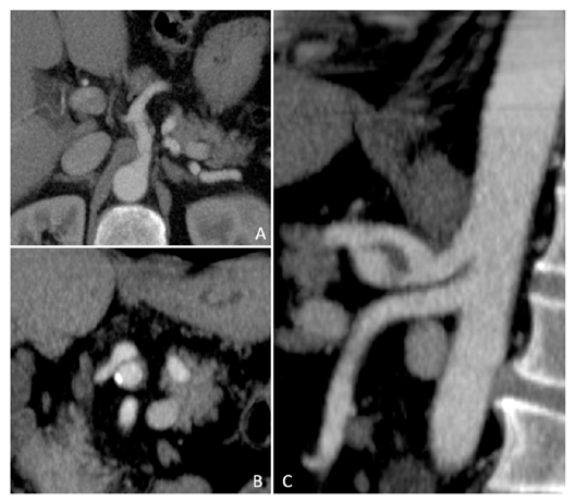
The definitive diagnosis is typically established by computed tomography angiography (Figure 1).
Treatment
In the absence of complications, most of the patients can be safely treated conservatively in an acute setting with food withdrawal, pain management, rigorous blood pressure control, antiplatelet agents, anticoagulation, and initially close clinical and imaging follow-up. Although recent literature suggests favorable outcomes after medical treatment, no established optimal medical treatment exists due to the rarity of this disease. The long-term prognosis of this disease also remains unknown.4
Surgical or endovascular management is reserved for the relatively exceptional cases of hemodynamic instability, persistent abdominal pain, progression of the dissection, end-organ ischemia, aneurysm/pseudoaneurysm degeneration, and rupture.
Endovascular approach has been usually the preferred initial approach. Several endovascular techniques have been described, depending on the anatomy of the dissection and its extension.7 Stenting is the most commonly applied treatment. Either bare metal stents and covered stents have been used. The latter are obviously preferred when aneurysm/pseudoaneurysm degeneration or rupture occurs. In both cases, self-expandable platforms should be preferred as they are much less aggressive to the arterial wall during deployment and their intrinsic outward force allows more smooth and regular apposition to the arterial wall along the artery, reducing further progression of the dissection.
Yet, in some clinical settings, embolization can be the option of choice. In those circumstances, coils should be preferred over plugs because the latter may induce an additional outward radial force that can lead to rupture of the weakened and actively expanding dissected artery. In very rare cases, both stenting and embolization may need to be considered; for instance, when the dissection involves both common hepatic and splenic arteries, embolization of the splenic artery and the deployment of a stent graft from the celiac to the common hepatic artery may need to be performed.
If endovascular treatment fails or is not possible, open surgery should be the next step, usually performing a graft interposition or bypass, although other procedures such as intimectomy, thrombectomy, or patch angioplasty has also been described, depending on the local anatomical situation.7
Follow-up
An aneurysmal degeneration can occur in the years following a spontaneous isolated visceral artery dissection as a result of the increased fragility of the arterial wall.7 The choice of imaging technique (computed tomography angiography, magnetic resonance angiography or ultrasound) will depend on the initial presentation, its evolution, and local expertise, even though direct comparison can be difficult to establish when different methods are performed. Since there are no studies with systematic and sufficiently long follow-up to determine the natural course after a spontaneous isolated visceral artery dissection, a follow-up imaging algorithm is challenging to establish.
Conclusion
Spontaneous isolated celiac and splenic artery dissection are very rare with less than 50 cases described so far. Even if they are associated with several possible risk factors, their definitive etiology remains unknown. The clinical presentation is quite variable ad most of the patients can be handled medically. However, endovascular or open surgery should be performed in case of complications. Careful follow-up, particularly in those patients treated conservatively, appears to be recommended.

