


 Inglês (pdf)
 Artigo em XML
 Referências do artigo
 Enviar este artigo por email
 Citado por SciELO
  Similares em
SciELO
Similares em
SciELO 
 Permalink
INTRODUCTION
An odontogenic cutaneous fistula (OCF) is one of the many consequences of chronic dental infection and it is defined as an abnormal communication between the face and the oral cavity, mostly due to a dental lesion.1 It is a rare and often misdiagnosed condition, leading inappropriate treatment and recurrence.
The causes include caries, pulp infection, periapical/ periodontal diseases, root fracture, and chemical/mechanical trauma.2 The average age of onset of OCF is 49 years, being slightly more frequent in male patients, usually with poor oral
hygiene.3
We present an illustrative case to draw the dermatologist’s attention to the importance of examining the oral cavity when facing perioral skin lesions, and considering a fistulous path secondary to odontogenic infectious processes.
CASE REPORT
A 32 years-old female patient, unemployed, sought dermatological care after referral from her family physician due to a mildly painful facial lesion present for more than 1 year, which had began as a small erythematous papule in the right nasogenian sulcus that rapidly progressed to a pustule, exudation and subsequent growth with intermittent episodes of local inflammation and pain, but no fever. A surgical excision was performed with immediate local recurrence.
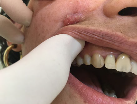
On dermatological observation there was an erythematous 1.2 cm nodule with a smooth and slightly shiny surface covered with thin yellowish crust that was mildly painful on palpation.
On the oral cavity the upper right canine tooth (tooth 13), which had had an extensive resin restoration on the mesial face, was clearly darker compared to the others (Fig.s 1 e 2). She mentioned adequate dental hygiene habits, but no dental consultation for more than 3 years.
Panoramic radiography was normal, but a facial computed tomography (CT) scan showed a periapical lucidity of the root of the upper right canine and an area of soft tissue density near this dental element, with a fistulous path that extended to the skin, confirming the diagnosis of an odontogenic cutaneous fistula. After endodontic treatment and surgical excision of the remaining skin lesion there was no further recurrence. Histopathology showed only a chronic non-granulomatous inflammatory process.
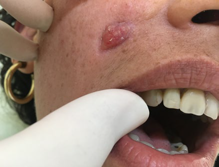
Figure 1 Lesão nodular eritematosa no sulco naso-geniano D, com alguma retração de tecido circundante
DISCUSSION
Misdiagnosis and inappropriate treatment are common for odontogenic cutaneous fistulas due to their rarity and lack of dental symptoms. The variability of morphological presentations and locations of the cutaneous lesions coupled with the lack of knowledge that such a condition can have a dental etiology generally leads to the misdiagnoses by surgeons and dermatologists, leading to unnecessary antibiotic and surgical therapies.1 Approximately 50% of the affected patients undergo multiple ineffective treatments (incision, drainage, and long-term antibiotics), due to improper diagnosis and lack of treatment of the dental infection.4
The cutaneous portion of odontogenic sinus is typically a nontender, erythematous nodule or a small nodulocystic lesion but ulceration, dyschromia, minor dimples, and other features may occur.5 Cords of tissue connecting the skin to the maxilla or mandible can be palpable and a pus-like discharge through the sinus opening may occur upon squeezing the cord. Perilesional skin can be slightly retracted.5
From a topographical point of view, periapical abscesses of the maxillary teeth can produce a cutaneous fistula that involves the cheeks, the nasolabial region, as in the present case, and even the inner canthus of the eye.3,5and the cutaneous fistula from odontogenic processes of the mandibula occur in the submandibular or submental region. More than one dental piece may be involved, more frequently in mandibular molar teeth (80%) than in maxillary teeth (20%).3
The absence of pain in most patients along with previous treatment attempts with incision and drainage, progressing to scarring and atrophic lesions can contribute to the delaying the correct diagnosis.3,4
The diagnosis is based on clinical suspicion and confirmed by a panoramic X-ray or especially by a periapical dental film, which show the radiolucency of the offending teeth.
The panoramic radiograph can reveal partial alveolar bone loss due to pulpitis and the presence of a periapical tooth abscess. CT scan, which greatly contributes to the evaluation of the cause and extent of infection, show bony changes such as an interruption or thinning of the alveolar cortical plate that surrounds the causal tooth, soft tissue changes such as thickening of the muscles and disappearance of the fat layer between the muscles, and paranasal sinus.5,6
Typically, the absence of prompt diagnosis and treatment results in pulp necrosis and the spread of infection beyond the affected tooth into the periradicular tissues, which often lead to an apical periodontitis. The chronic process slowly evolves through the cancellous alveolar bone, following the path of least resistance until it perforates the cortical plate of the mandible, spreading to the surrounding soft tissues and erupting on the skin.1
Dental treatment is the mainstay of therapy: endodontic treatment or surgical root canals and apicoectomies for restorable teeth and extraction of non-vital teeth can eliminate infection. The sinus tract will typically resolve within 5 to 20 days, while minimal scarring and hyperpigmentation can remain.
Surgical revision for scar tissue can be delayed.3,5,6
Recurrence will most likely occur in case of lesional excision without suitable treatment of the affected teeth.3,5
Possible complications associated with a untreated OCF are osteomyelitis of the jaw, Ludwig's angina, mediastinitis and even septicemia, which can end up compromising the patient's life.3
With this case, we want to draw attention of the dermatologist and even the general practitioner to odontogenic fistulas, especially extra-oral ones with cutaneous involvement, emphasizing the importance of a careful history, meticulous examination, adequate confirmation through imaging and treatment by dental technicians in a multidisciplinary approach.

