Inglês (pdf)
 Artigo em XML
 Referências do artigo
 Enviar este artigo por email
 Citado por SciELO
  Similares em
SciELO
Similares em
SciELO 
 Permalink
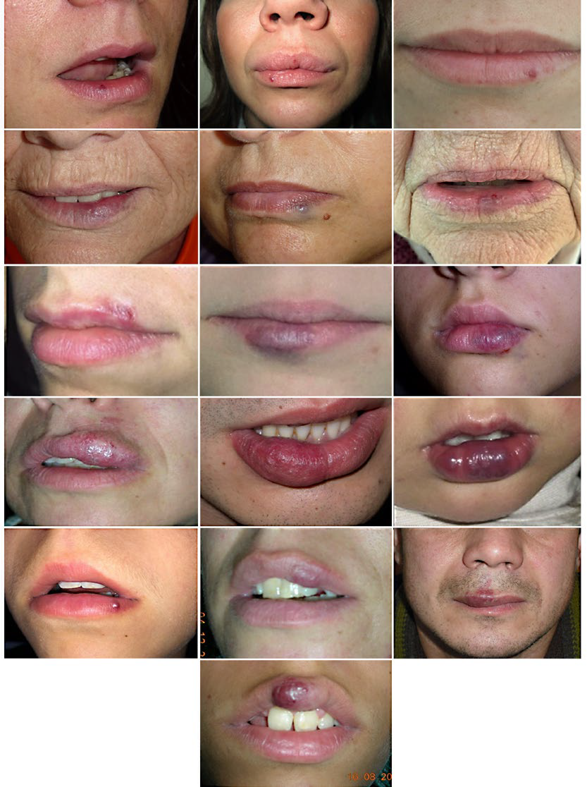
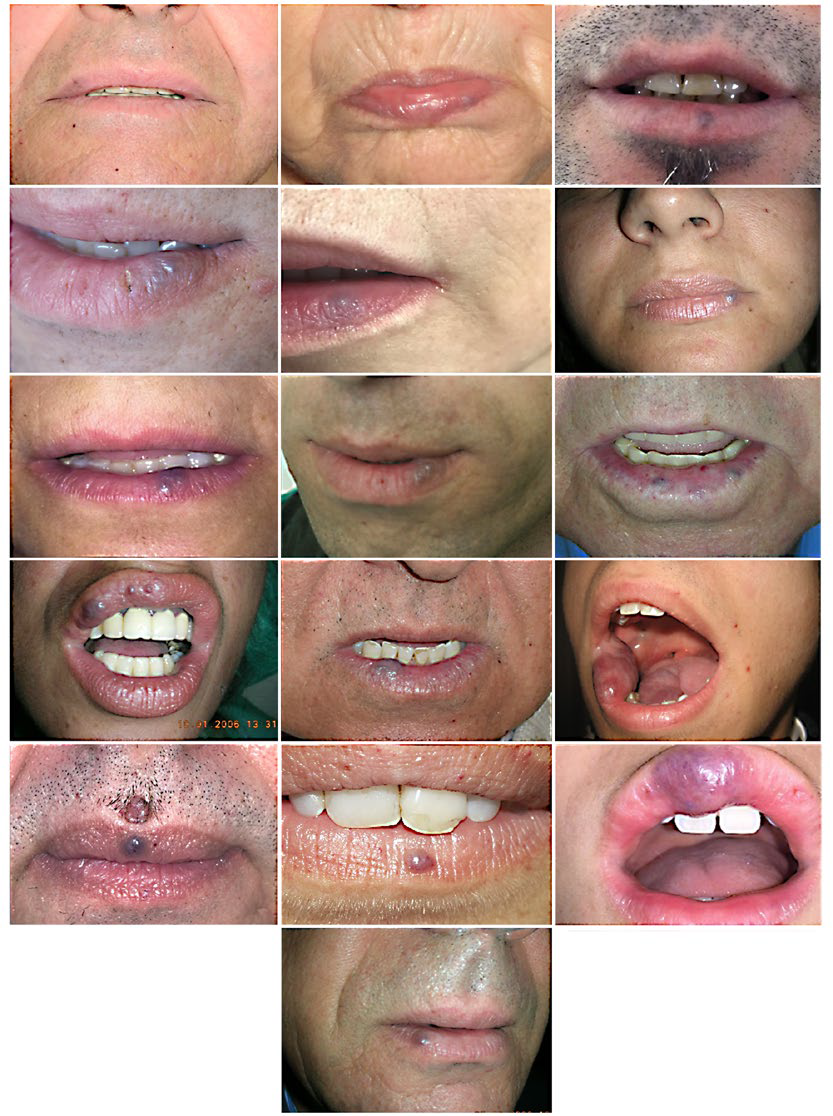
Vascular malformations are localized defects of vascular morphogenesis, likely caused by dysfunction in pathways regulating the angiogenesis. According to the classification system adopted by the International Society for the Study of Vascular Anomalies (ISSVA), vascular malformations are categorized as capillary, venous, lymphatic and arteriovenous, depending on the predominant type(s) of anomalous vessels. Categorizing a lesion as a capillary malformations (CM), venous malformations (VM), lymphatic malformations (LM), arteriovenous malformations (AVM), or combined lesion is important, since the prognosis and the treatment differ depending on the type(s) of vessels involved.1,2However, small vascular malformations of the lip can pose difficult diagnostic challenges, due to their parallel natural history and clinical similarities (Fig.s 1,2).
The detection of some specific features may facilitate the diagnosis and allow the distinction between VM and AVM.
Venous malformations typically present as slow-growing, soft, compressible, blue to violaceous nodules, that are usually asymptomatic and have tendency to fill with Valsalva maneuver or dependent position. Dermoscopic assessment of this lesions commonly shows the presence of clods and a structureless pattern that may vary in color.3
In contrast, arteriovenous malformations are characterized by na erythematous, firm, pulsatile swelling that may demonstrate a palpable thrill and tendency to bleed. On occasions, rapid expansion can occur, especially following trauma, infection and hormonal changes. Arteriovenous malformations are typified dermoscopically by blue-red lacunae, milky red areas and winding vessels of several thicknesses. However, when clinical doubt persists, doppler ultrasonography may indicate the definitive diagnosis by differentiating low from high vascular flow.
.