


 English (pdf)
 Article in xml format
 Article references
 Send this article by e-mail
 Cited by SciELO
  Similars in
SciELO
Similars in
SciELO 
 Permalink
Dear Editor
We read with great interest the article “Mechanic’s Hands and Drug Eruption to Hydroxycholoroquine: Precious Diagnosis Tools”.1 As a result we report a case of dermatomyositis with a different clinical presentation that will aggregate value to the clinical findings on this disease.
A 78-year-old man, with a history of polycystic kidney disease (G3a KDIGO) and hypertension, presented to his primary care physician with a facial erythema and periocular edema (heliotrope rash) and a violaceous macular nonpalpable erythema on the neck, chest and back with intense pruritus after three days of sun exposure (Fig. 1A). Oral deflazacorte (30 mg/day) was prescribed for 10 days, without clinical improvement. Thus, the patient presented to the emergency department with worsening of cutaneous symptoms, myalgias and symmetric muscle weakness with proximal predominance, asthenia and fever (39ºC, tympanic). He denied dyspnea, dysphagia, arthralgia and Raynaud’s phenomenon and infection was excluded. He also had a flagellate erythema in the abdomen (Fig. 1B), erythematous papules on the back of hands over metacarpophalangeal and interphalangeal joints, erythema in the nail folds and purpuric lesions on the finger
pulps and nail cuticula and hyperkeratotic and fissured lesions on fingers and palms (Fig. 1C). Laboratory workup revealed elevated muscle enzymes (creatine kinase: 3172U/L, aspartate aminotransferase: 219U/L, alanine aminotransferase:
85U/L, lactate dehydrogenase: 420U/L) and worsening of renal function (urea: 190 mg/dL, creatinine: 2.41 mg/dL).
Capillaroscopy of nail bed showed capillary loss (Fig. 1D). Electromyography of deltoid muscle showed a low-amplitude and short-duration polyphasic motor unit potential, consistent with myopathy. Antinuclear antibodies (ANA) and antibodies to Transcriptional intermediary factor 1 gamma (TIF-1γ) were positive. The remaining autoimmunity profile was negative.
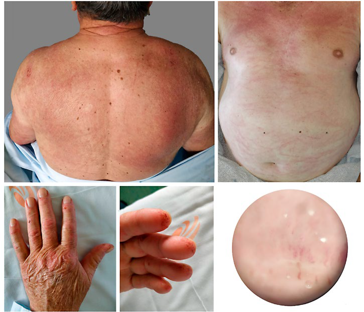
Figure 1 Clinical presentation and capillaroscopy: (A) Itchy violaceous macular non palpable erythema on the back (photodistributed poikiloderma). (B) Violaceous macular nonpalpable erythema on the neck and chest (V sign) and flagellate erythema in the abdomen. (C) Erythematous papules on the back of hands (Gottron´s papules), periungueal erythema and purpura in the nail cuticula and finger pulps. (D) Capillaroscopy: loss of capillaries in the proximal nail fold
With the diagnosis of dermatomyositis, prednisolone (1 mg/ kg/day) was started. He was asymptomatic at 4 weeks with normalization of muscle enzymes at 6-weeks. Glucocorticoids were progressively reduced. A full-body computed tomography scan excluded an occult tumor.
Dermatomyositis is a rare autoimmune condition that occurs in children and adults and affects the skin and muscles.2
Apart from classic rashes, less frequent cutaneous lesions as flagellate erythema are also described,3-6 as in this case. Muscle weakness, increased muscle enzymes and abnormal electromyography allow the diagnosis of dermatomyositis, even without muscle biopsy, in the light of the latest European League Against Rheumatism (EULAR) criteria.3,4Myositis specific autoantibodies represent a complement to the diagnosis and a guide for treatment and follow-up.5,6Among these, Anti-transcriptional intermediary factor-1γ (TIF-1γ) autoantibodies correlates with the presence or development of neoplasia in the future, which implies looking for occult neoplasia and maintaining a closer follow-up.5,6Prednisolone (1 mg/kg/day) is the first line treatment and symptom relief is expected within 4 weeks, after which a slow reduction is recommended,2 but methotrexate or azathioprine may be used as a second-line or as a steroid sparing agent.2

