INTRODUCTION
Coronavirus are encapsulated RNA viruses belonging to the Coronaviridae family and to the order Nidovirales, widely distributed among humans and other mammals.1 In 2019, a series of pneumonia cases of undetermined cause with clinical involvement similar to viral pneumonias appeared in China.2 Sequenced analyzes of the patients' lower respiratory tract identified a new version of the virus, the new coronavirus 2019 (nCov-19).1-3
The disease became known as “severe acute respiratory syndrome coronavirus 2” (SARS-CoV-2) and gained global proportions, configuring a pandemic, given its high transmission capacity between humans. Patients infected with SARS -CoV-2 may be oligosymptomatic or even asymptomatic, but a large part of those affected develop symptoms such as fever, cough, anosmia, dysgeusia, myalgia and headache, in addition to lung disease, which can cause respiratory failure and respiratory syndrome acute severe (SRAG), which generates the need for hospitalization and intensive care.1,3,4Besides involvement of the digestive, cardiovascular and nervous system, this virus also generates nonspecific changes in the skin, and as far as we know they are not fully related to prognosis.5-11COVID-19 pandemic fatalities are rare before puberty (<10 years of age), and the vulnerability of males to severe disease has been constantly reported over the past months of pandemic.6
Brazil currently has more than 4,5 million confirmed cases and almost 137 thousand deaths, occupying the second place in the world in number of cases, behind only the United States.7
In this article we will present a series of 19 cases of Brazilian patients infected with SARS-CoV-2 and who developed dermatological lesions. We will describe the polymorphism of the skin lesions and make a correlation in relation to the chronology of the infection, aiming at whether the dermatological lesions occurred before, during, after or not related to the systemic symptoms due to this infection. We hope to contribute to a better understanding of dermatological involvement and to warn about the possibility of the occurrence of dermatological lesions, as a prodromal sign or unique symptom of COVID-19.
MATERIAL AND METHODS
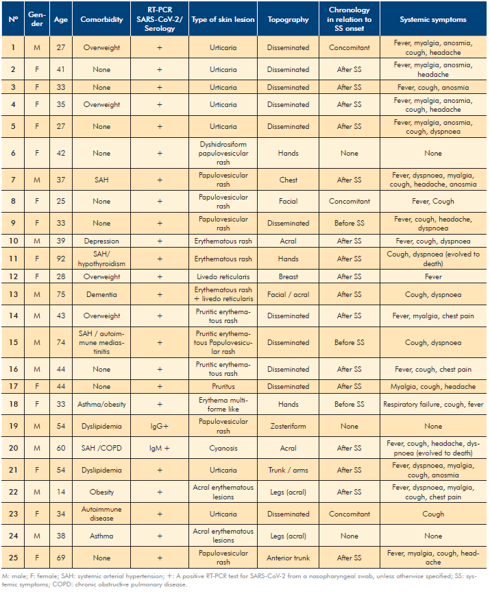
An observational study was carried out of patients seen at outpatient dermatology consultations and of patients hospitalized for clinical management in Rio de Janeiro state, Brazil. We evaluated 25 cases of patients affected by SARS-CoV-2 who had dermatological lesions. We analyzed objectively their data such as sex, age, comorbidities, laboratory tests related to SARS-CoV-2 infection, contact with confirmed cases, type of dermatological lesions, their topography, chronology according to systemic symptoms and which systemic symptoms were presented.
RESULTS
We analyzed data from 25 patients, aged between 14 and 92 years old. Of this population, 9 patients did not have comorbidities, 3 were overweight, 4 had systemic arterial hypertension, one of whom was also affected by hypothyroidism, another by chronic obstructive pulmonary disease (COPD) and another onde by autoimmune mediastinitis; 1 dementia, 1 depression, 1 obesity, 1 asthma, 1 obesity and asthma, 2 dyslipidemia and 1 autoimmune disease.
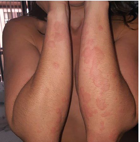
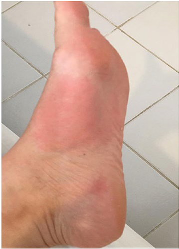
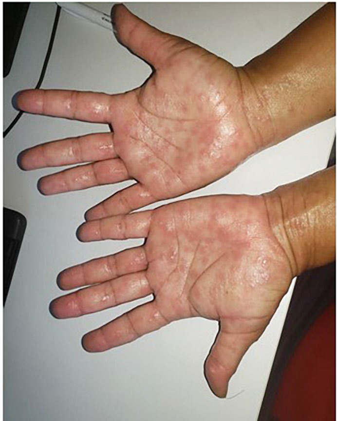
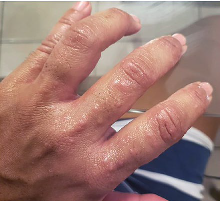
Among all patients, 23 had positive reverse transcriptase- polymerase chain reaction (RT-PCR) for SARS-CoV-2, one had positive IgM serology and another one had positive IgG serology. They presented skin lesions of the following types: urticaria / annular urticarial rash (Fig. 1), pruritic erythematous rash (Fig. 2), erythema multiforme like rash (Fig. 3), pruritic erythematous papulovesicular rash (Fig. 4), livedo reticularis (Fig. 5), dyshidrosiform papulovesicular rash (Fig. 6), cyanosis and acral erythematous lesions.
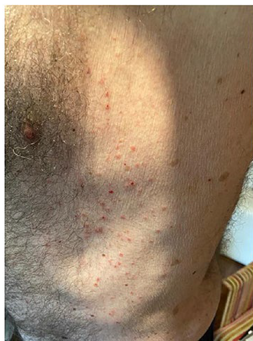
Figure 4 Pruritic erythematous papulovesicular rash that preceded the onset of flu-like symptoms (Patient Nº 15).
Three patients did not present any systemic symptoms, sixteen had skin lesions after the symptoms, three during the symptoms and three had skin lesions preceding the systemic symptoms. Patients number 11 and 20 died due to COVID-19 infection. Details are in Table 1.
DISCUSSION
Brazil has presented alarming and growing data on infection and deaths from SARS-CoV-2. There are more than 4 500 000 confirmed cases and more than 137 000 deaths ranking second in the world ranking of infected people.10
In Lecco, Italy, an observational study was conducted with a sample of patients infected with SARS-CoV-2. From the collected data (88 patients), 18 patients (20.4%) developed cutaneous manifestations. Eight patients developed cutaneous involvement at the onset, 10 patients after the hospitalization.8
Skin involvement was observed in 0.2%-1.2% in 1099 patients with laboratory-confirmed COVID-19 from 552 hospitals in 30 provinces, autonomous regions, and municipalities in mainland China through January 2020.9
According to the Brazilian Ministry of Health, Rio de Janeiro has an alarming number of confirmed cases, with a total of 277 439 infected.10 However we did not find any records of the frequency of cutaneous involvement in infected patients.
Spanish researchers conducted a study in which data were collected from 375 patients infected with SARS-CoV-2 who had cutaneous manifestations.
Among these patients 19% presented pseudo-chilblain lesions, 9% vesicular lesions, 19% urticarial lesions, 47% maculopapular lesions and 6% livedo / necrosis lesions.12
The chronology of skyn manifestations has also been described.
Among pseudo-chilblain lesions, 7% occurred before, 34% during and 59% after systemic symptoms. Among vesicular lesions, 15% occurred before, 56% during and 29% after systemic symptoms. Of the urticaria lesions, 4% before, 61% during and 35% after symptoms. The maculopapular lesions occurred in 5% before, 61% during and 34% after symptoms.
Livedo / necrosis lesions occurred in the proportion of 5% before, 86% during and 10% after systemic symptoms.12
In our study we evaluated 25 patients infected with SARS-CoV-2 who had cutaneous lesions. The frequency of the types of skin lesions presented was 28% for urticarial lesions, 16% erythematous rash, 4% erythema multiforme like rash, 24% papulovesicular rash, 8% livedo reticularis, 4% dyshidrosiform papulovesicular rash, 4% cyanosis, 4% pruritus and 8% for acral erythematous lesions.
Concerning the chronology of skin involvement 12% had cutaneous lesions before the symptoms, 12% at the same time and 64% after onset of systemic symptoms, wherea 12% of patients did not present any systemic symptoms related to COVID-19.
CONCLUSION
It is of great importance that the dermatologist be aware of the chronology of cutaneous involvement of COVID-19.
Few cases may present with skin lesions as an isolated symptom, while other skin signs may precede systemic involvement.
This clinical perception may guarantee a more effective and safer approach, both in terms of preventing the spread of the virus, by isolating these patients with sugestive epidemiological data, as well as detailed clinical follow-up of patients, with greater vigilance regarding the development of severe symptoms. Although the number of cases on the world stage seems to be regressing, infection by SARS-CoV-2 will be part of the dermatologist's routine. As long as we do not have a widely available vaccine and the pandemic takes on an endemic profile, we must be attentive to these manifestations, not only for the proper diagnosis, indication of patient isolation, as well as all the necessary biosafety procedures in dermatological clinics.


