SciELO - Scientific Electronic Library Online

 
vol.78 issue4COVID-19 and Cutaneous Disorders: What’s Being Reported? A Meta-Analysis from Observational Studies and Case ReportsPhototherapy in Pediatric Population: Particularities and Indications author indexsubject indexarticles search
Home Pagealphabetic serial listing  

Services on Demand

Journal

Article

Indicators

Related links

  • Have no similar articlesSimilars in SciELO

Share


Revista da Sociedade Portuguesa de Dermatologia e Venereologia

Print version ISSN 2182-2395On-line version ISSN 2182-2409

Rev Soc Port Dermatol Venereol vol.78 no.4 Lisboa Dec. 2020  Epub Sep 10, 2021

https://doi.org/10.29021/spdv.78.4.1270 

Artigo de Revisão

Cutaneous Squamous Cell Carcinoma in Solid Organ Transplant Patients

Carcinoma Espinocelular em Doentes com Transplante de Órgão-Sólido

1Serviço de Dermatologia, Hospital de Egas Moniz, Centro Hospitalar Lisboa Ocidental, Lisboa, Portugal

2Serviço de Dermatologia, Hospital de Santa Maria, Centro Hospitalar Universitário Lisboa Norte, Lisboa, Portugal

3Clínica Universitária de Dermatologia de Lisboa, Faculdade de Medicina, Universidade de Lisboa. Lisboa, Portugal


ABSTRACT

Solid organ transplant recipients (SOTR) are more likely to develop cutaneous squamous cell carcinoma (CSCC), compared to the general population. This increased incidence is due to several factors including chronic ultraviolet radiation exposure, human papillomavirus (HPV) infection, immunosuppressive medication, among others. Primary prevention is crucial, because not only are cutaneous squamous cell carcinomas more frequent in SOTR, but they are also more aggressive and have therefore a worse prognosis. Newer immunosuppressive drugs are associated with a smaller risk of developing CSCC, but they can have more adverse events, prompting patients to abandon therapy. Concerning treatment options for CSCC, they range from surgery, radiotherapy, and systemic therapy, although there are few studies in SOTR concerning the use of immunotherapy and epidermal growth factor receptor (EGFR) inhibitors.

KEYWORDS: Carcinoma, Squamous Cell; Immunosuppression/adverse effects; Skin Neoplasms; Transplant Recipients.

RESUMO

Os doentes com transplante de órgão-sólido têm maior incidência de carcinoma espinocelular (CEC) do que a população em geral. Esta maior incidência deve-se a diversos fatores, entre os quais, exposição crónica à radiação ultravioleta, infeção por papiloma vírus humano, fármacos imunossupressores, entre outros. A prevenção primária é crucial, porque além dos CEC serem mais frequentes nos doentes transplantados, são também mais agressivos e têm, portanto, um prognóstico pior. Os novos fármacos imunossupressores estão associados com um risco menor de desenvolvimento de CEC, no entanto, podem ter mais efeitos adversos, levando ao abandono da terapêutica. Em relação às opções terapêuticas para CEC, estas variam entre cirurgia, radioterapia e terapêutica sistémica, apesar de existirem poucos estudos em doentes com transplante de órgão-sólidos, em relação à imunoterapia e inibidores do recetor do fator de crescimento epidérmico.

PALAVRAS-CHAVE: Carcinoma de Células Escamosas; Imunossupressão/efeitos adversos; Neoplasias da Pele; Transplantados

INTRODUCTION

After basal cell carcinoma (BCC), the second most common malignancy is cutaneous squamous cell carcinoma (CSCC).1,2Its incidence has continued to rise, with estimates that it has increased from 50% to 300% in the last thirty years, and that in the next decade, its incidence is predicted to double.3-5

For Caucasians, the risk of developing a CSCC in a lifetime is 7% to 11%.6 The risk is much higher if we take into account, solid organ transplant recipients (SOTR), who have a 65-250-fold greater risk.7,8In contrast, the incidence of basal cell carcinoma is only 6-10 fold higher in SOTR.9,10

Consequently, instead of the normal BCC/CSCC ratio of 4:1 in the general population, this ratio has been reported as being inverted in SOTR, with CSCC being four times more common than BCC.8

In comparison with renal or liver transplant recipients, heart or lung transplant recipients have been found to have an even higher incidence of CSCC.10 This incidence is probably due to more intensive immunosuppressive regimens required after heart or lung transplants as well as the older age of transplanted patients.9

For localized CSCC, the ten-year-survival after surgery surpasses 90%, but these tumors can be locally invasive or even metastatic, with rates of lymph node metastases of about 4% and mortality rates of almost 2%.11,12In SOTR, not only are CSCC more frequent, but they are also more aggressive, with greater tumor depth, higher rates of recurrence and higher rates of local and distant invasion.13,14

PORTUGUESE REALITY

In the last 10 years, four reviews about cutaneous malignancies in SOTR were published.15-18 Data about these studies is summarized in Table 1. In these publications, the number of transplant recipients ranged from 127-319 patients, with a mean age of 50.7-63.5 years and a predominance of male patients (60.9%-88.9%). The number of patients who developed CSCC fluctuated between 10-62, with an average time elapsed since transplant of 5-8 years.

Contrary to the inverted ratio 4:1 of BCC and CSCC described in several anglo-saxonic publications,8 in the Portuguese series this inversion has not been verified.15-18

Instead, ratios of 1:1 have been found between BCC and CSCC. This is similar to other Southern European reports, such as those from Spain19,20and Italy.21 These ratios are attributed to genetic differences as well as sun exposure habits.16,18

Table 1 Summary of Portuguese revisions about CSCC in SOTR, in the last 10 years 

Publications Operation year Number of transplant recipients Mean age+ Male gender Number of patients that developed CSCC Time interval from transplant until CSCC* Ratio BCC: CSCC
Fernandes et al15 2000-2010 319 (180 renal and 230 hepatic) 60.5 - renal 63.5 - hepatic 83.3% - renal 88.9% - hepatic 18 55.2 - renal 36 - hepatic 1:1.3 - renal 3.5:1- hepatic
Borges-Costa et al16 2010-2011 127 renal 53 67% 10 96 1:1
Pinho et al17 2004-2013 288 renal 61.9 66% 62 64 1:1
Garrido et al18 2008-2014 197 renal 50.7 60.9% 15 Not specified 1.1:1

RISK FACTORS

The risk of developing a CSCC at 10 years post-transplant is estimated to be 10%-27% and at 20 years post-transplant is 40%-60%.9

The most contributing factor is ultraviolet (UV) radiation, explaining why the majority of CSCC occur in sun-exposed locations (Fig.s 1 and 2).22Additional contributing factors are advanced age, male gender, skin phototype I or II, infection with beta human papillomaviruses (HPV), genetic factors and the immunosuppressive medication that is needed to maintain the transplanted organ.16,17,23

UV radiation exposure causes mutations in tumor suppressor genes, such as TP53 and NOTCH, and oncogenes, like HRAS, NRAS and KRAS.24 Even sun-exposed skin, without clinical abnormalities, has a similar amount of mutations when compared with oropharyngeal squamous cell carcinomas, although it has a 10-fold lower mutations amount than that reported for CSCC.24

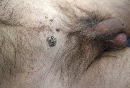
HPV have also been implicated in the pathogenesis of CSCC (Fig. 3).25,26It is well established the role of alpha-genus HPV, such as HPV 16 and HPV 18, in the development of anogenital carcinoma, and they are also involved in the formation of head and neck CSCC and periungual CSCC.27

Beta-genus HPV, like HPV5, HPV8 and HPV9 can be present in the skin of healthy people, but their levels rise with immunosuppression.28,29Their presence has been found in almost 90% of CSCC in SOTR and has been associated with twice the risk of CSCC in SOTR.28,29

A supporting finding for the implication of HPV in CSCC is that actinic keratosis, which are precursors of CSCC, also have an elevated number of Beta-HPV copies.30

The mechanism of action that is hypothesized in the genesis of anogenital CSCC involves the inhibition of TP53 and retinoblastoma protein by the HPV’s oncoproteins E6 and E7.31 Beta-HPV may allow the accumulation of UVR induced mutations, through several mechanisms, for instance, degradation of Bak (involved in cellular apoptosis) and inhibition of Notch tumor suppression.24,27,28

Regardless of these findings, there are some studies in which HPV is found in low or absent activity in CSCC, and this led to the “hit-and-run” hypothesis, that states that Beta-HPV are involved in the formation of the tumor but they are not necessary for its maintenance, contrary to alfa-HPV in anogenital SCC.27,28,31

Another important risk factor is the immunosuppressive medication used for the organ maintenance.32 Sirolimus has been demonstrated to reduce the number of CSCC in comparison to calcineurin inhibitors tacrolimus and cyclosporine.33 The benefit of switching to sirolimus is supposed to be greatest after the development of one CSCC and negligible when there are multiple prior CSCC. Therefore, it has been suggested to switch from calcineurin inhibitors to sirolimus after the first post-transplant CSCC.3,8,13Despite this, sirolimus has more serious adverse events per patient, prompting more patients to discontinue treatment, and in a meta-analysis it has been associated with an increased risk of death, although this remains controversial.33,34

Azathioprine, that acts as a photosensitizer to UVA, is also responsible for an increased risk of CSCC.16Mycophenolate mofetil seems to be associated with lesser risk and should be considered as an alternative option to azathioprine.35

All the factors cited above, act synergistically and as a result of an interaction there can be development and progression of CSCC in solid organ transplant recipients.24

Figure 1 Cutaneous squamous cell carcinoma on the hand of a renal transplant recipient. 

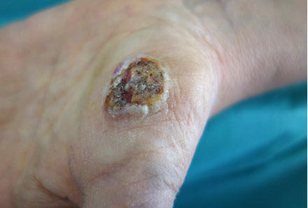
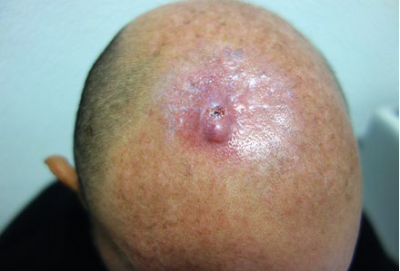
Figure 2 Recurrence of cutaneous squamous cell carcinoma on the head of a renal transplant recipient. Squamous cell carcinoma is more frequent in sun-exposed locations. 

Figure 3 Cutaneous squamous cell carcinoma on the genital area of a male patient. HPV infection is a contributing factor in th e development of CSCC, especially on this area. 

PREVENTION

Since SOTR have a higher incidence of CSCC and these are more aggressive, with an increased risk of metastasis of 5%-7%, skin cancer prevention is essential.36

Given that the most important risk factor is UV radiation, emphasis on sunscreen use and sun protection must be made.9

Before transplant, patients should have a full-body skin examination, with a subsequent periodic assessment depending on previous or concomitant skin premalignant and malignant lesions.9 (Table 2). An annual screening is suggested for patients without pre-malignant or malignant lesions. Patients with actinic keratosis or one prior nonmelanoma skin cancer should be evaluated at 3-6 months and if a high-risk CSCC is found the follow-up should be every 3 months. In patients presenting with a metastatic squamous cell carcinoma, an examination after 1-3 months is advised.9

Table 2 Recommended screening interval, according to patient’s risk factors 

Risk factors Periodic assessment
Absence of pre-malignant / malignant lesions Annual screening
Pre-malignant lesions/ One prior nonmelanoma skin cancer 3-6 months
Multiple prior nonmelanoma skin cancers/ High-risk CSCC 3 months
Metastatic CSCC 1-3 months

STAGING

There are three main staging systems used for staging CSCC: the American Joint Committee on Cancer (AJCC), the Union for International Cancer Control (UICC) and, more recently, the Brigham and Women’s Hospital Tumor staging system.13.22These staging systems have into account factors such as: tumor dimensions, perineural invasion, depth of invasion and poorly differentiated histology.13.22

Nevertheless, some criteria with prognostic influence, like tumor location and immunosuppression are not considered in any of the main staging systems, causing a potential underestimation of CSCC prognosis in SOTR.37

TREATMENT

Considering the aggressiveness of CSCC in SOTR, treatment of pre-malignant lesions should be sought out and if there is suspicion of transformation into malignant lesions, biopsy or excision should be performed.38

In terms of topical therapies for the treatment of actinic keratosis, there is a wide range of options available: diclofenac, 5-fluorouracil, imiquimod, laser, cryotherapy, among others.22,38

In single lesions, local destructive options, such as cryotherapy or electrocautery and curettage can be considered.

Clearance of 41% of lesions has been achieved with topical diclofenac and 62% clearance has been obtained with topical imiquimod.39,40Imiquimod is an immunomodulatory agent that has anti-viral and anti-tumor properties.41 It has been advised to limit its use to an area of 60-100 cm2, because albeit rare, imiquimod has been linked to induction of systemic immunostimulation.42 However some studies have demonstrated that the actual systemic absorption of topical imiquimod is minimal and hundreds of patients have been treated without graft rejection.42 For areas with extensive lesions, photodynamic therapy should be considered, and has been proven to be more effective than imiquimod.38,43

For CSCC, surgery is the gold standard treatment, not only because it allows tumor removal but also histologic verification.44 The recommended surgical margins depend on the size of the tumor, with margins of 0.4 cm being advocated for tumors inferior to 2 cm, and margins of 0.6 cm for tumors larger than 2 cm.45

For high risk locations (scalp, ears, eyelids, nose), Mohs micrographic surgery is recommended, but if this is not possible and standard excision is performed, avoidance of significant tissue rearrangement is necessary, before confirming histologic margins are free of tumor.44-46Although radiotherapy for CSCC has declined, it can be a viable alternative when surgical options are not possible, particularly for primary tumors.44 An overall control rate was achieved for 89% of previously untreated CSCC and 68% for ecurrent CSCC.47 Radiotherapy is also used as an adjuvant treatment of surgery, for CSCC with perineural invasion, although it has not been established that it improves survival.48,49

In locally advanced or metastatic CSCC, if surgery or radiotherapy are not feasible, systemic therapy with cytotoxic agents (platinum, interferon-alpha, 5-fluouracil and taxanes), epidermal growth factor receptor (EGFR) inhibitors and immunotherapy can be used.22 However, in SOTR there are few studies evaluating the safety and efficacy of these treatments.22

Depending on the treatment combination used, response rates with chemotherapy have ranged from 14% to 86%, with cisplatin and 5-fluouracil obtaining the best results.50

Complete remission was achieved in 43% of patients treated in one series, however side effects of these treatments should be considered.50

The EGFR inhibitors, cetuximab and panitumumab have shown some response against advanced CSCC, with response rates of 27%, and 31%, respectively, evaluated in immunocompetent individuals.49-52These can represent valid options for tumors expressing high levels of EGFR, however in SOTR there are few studies available.50,51

Immunotherapy with anti-PD-1 monoclonal antibody, specifically cemiplimab, was approved in 2018 for the treatment of advanced CSCC.53 The response rate was 47% with an overall survival of 81%.53 Despite these high response rates, in SOTR, there is a risk of organ rejection with checkpoint inhibitors, that is almost 50% within a month of therapy.22

CHEMOPROPHYLAXIS

For patients who develop more than 5 CSCC/per year, systemic retinoid therapy should be considered.54 Acitretin, a systemic retinoid, has reduced the incidence of actinic keratosis and CSCC in SOTR.55 Starting with a low dose of 10mg/daily is recommended, with maximum doses of 30 mg/ daily, in order to prevent patient loss of compliance, due to adverse events.56

However, there are several contraindications for their use, like pregnancy, severe hyperlipidemia, and a rebound effect when therapy is discontinued.57

For this reason, therapy with systemic retinoids should be carefully weighted, as patients can experience a high number of CSCC in a short period of time.55,56Another option that has been studied for chemoprophylaxis, is oral capecitabine, a pro-drug of 5-fluorouracil, that has also shown to reduce the incidence of pre-malignant and malignant lesions after solid organ transplant, with less side effects than systemic retinoids.58

CONCLUSION

Cutaneous squamous cell carcinoma incidence has been rising and is predicted to continue to increase. These tumors are even more common and aggressive in solid organ transplant recipient patients. Ultraviolet radiation is the most important risk factor for the development of CSCC and so primary prevention must be implemented amongst patients. Immunosuppressive medication is necessary to prevent organ rejection but is also associated with an increased risk of developing skin malignancies. Particularly, old immunosuppressive drugs, such as, azathioprine, cyclosporine and tacrolimus have been linked to a higher incidence of CSCC.

Primary treatment option is surgical excision with surgical margins depending on tumor’s size. Radiotherapy can also be used as a therapeutic alternative. Chemoprophylaxis with acitretin should be considered for patients that develop more than 5 CSCC/per year.

REFERENCES

1. Rogers W, Weinstock MA, Feldman SR, Coldiron, BM. Incidence Estimate of Nonmelanoma Skin Cancer (Keratinocyte Carcinomas) in the U.S. Population, 2012. JAMA Dermatol. 2015; 151:1081-6. doi: 10.1001/jamadermatol.2015.1187. [ Links ]

2. Muzic JG, Schmitt R, Wright AC, Alniemi DT, Zubair AS, Baum CL, et al. Incidence and Trends of Basal Cell Carcinoma and Cutaneous Squamous Cell Carcinoma: A Population-Based Study in Olmsted County, Minnesota, 2000 to 2010. Mayo Clin. Proc. 2017; 92: 890-8. doi: 10.1016/j.mayocp.2017.02.015. [ Links ]

3. Corchado-Cobos R, García-Sancha N, González-Sarmiento R, Pérez-Losada J, Cañueto J. Cutaneous Squamous Cell Carcinoma: From Biology to Therapy. Int J Mol Sci. 2020; 21:2956. doi: 10.3390/ijms21082956. [ Links ]

4. Brougham ND, Tan ST. The incidence and risk factors of metastasis for cutaneous squamous cell carcinoma - Implications on the T-classification system. J Surg Oncol. 2014; 110: 876-82. doi: 10.1002/jso.23731. [ Links ]

5. Leiter U, Keim U, Eigentler T, Katalinic A, Martus P, Garbe C. Incidence, Mortality, and Trends of Nonmelanoma Skin Cancer in Germany. J Investig Dermatol. 2017; 137:1860-7. doi: 10.1016/j.jid.2017.04.020. [ Links ]

6. Miller DL, Weinstock MA. Nonmelanoma skin cancer in the United States: Incidence. J Am Acad Dermatol. 1994; 30:774-8. [ Links ]

7. Bangash HK, Colegio OR. Management of non-melanoma skin cancer in immunocompromised solid organ transplant recipients. Curr Treat Options Oncol. 2012; 13:354-76. doi: 10.1007/s11864-012-0195-3. [ Links ]

8. Euvrard S, Kanitakis J, Claudy A. Skin cancers after organ transplantation. N Engl J Med. 2003; 348:1681-91. [ Links ]

9. Zwald FO, Brown M. Skin cancer in solid organ transplant recipients: advances in therapy and management: part I. Epidemiology of skin cancer in solid organ transplant recipients. J Am Acad Dermatol. 2011; 65:253-61. doi: 10.1016/j.jaad.2010.11.062. [ Links ]

10. Krynitz B, Olsson H, Lundh B, Lindelof B, Edgren G, Smed KE. Risk of basal cell carcinoma in Swedish organ transplant recipients - a population -based study. Br J Dermatol. 2015; 174:95-103. doi: 10.1111/bjd.14153. [ Links ]

11. Varra V, Woody NM, Reddy C, Joshi NP, Geiger J, Lamarre, E.D, et al. Suboptimal Outcomes in Cutaneous Squamous Cell Cancer of the Head and Neck with Nodal Metastases. Anticancer Res. 2018; 38: 5825-30. doi: 10.21873/anticanres.12923. [ Links ]

12. Schmults CD, Karia PS, Carter JB, Han J, Qureshi A. Factors predictive of recurrence and death from cutaneous squamous cell carcinoma: A 10-year, single-institution cohort study. JAMA Dermatol. 2013; 149: 541-7. doi: 10.1001/jamadermatol.2013.2139. [ Links ]

13. Kim C, Cheng J, Colegio OR. Cutaneous squamous cell carcinomas in solid organ transplant recipients: emerging strategies for surveillance, staging, and treatment. Semin Oncol. 2016; 43:390-4. [ Links ]

14. Lott DG, Manz R, Koch C, Lorenz R. Aggressive behavior of non-melanotic skin cancers in solid organ transplant recipients. Transplantation. 2010; 90:683-7. [ Links ]

15. Fernandes S, Carrelha AS, Marques Pinto G, Nolasco F, Barroso E, Cardoso J. Patologia dermatológica em doentes transplantados hepáticos e renais referenciados à consulta de dermatologia e venereologia. Acta Med Port. 2013;26:555-63. [ Links ]

16. Borges-Costa J, Vasconcelos JP, Travassos AR, Guerra J, Santana A, Weigert A, et al. Cancro Cutâneo em Doentes com Transplante Renal: Incidência e Associações com Fatores Clínicos e Sociodemográficos. Acta Med Port. 2013;26:123-6. [ Links ]

17. Pinho A, Gouveia M, Cardoso JC, Xavier MM, Vieira R, Alves R. Non-melanoma skin cancer in portuguese kidney transplant recipients - incidence and risk factors. Na Bras Dermatol. 2016; 91:455-62. [ Links ]

18. Garrido PMC, Borges-Costa J. Skin disorders in renal transplant recipients: a retrospective study. An Bras Dermatol. 2017; 92:638-41. [ Links ]

19. Marcén R, Pascual J, Tato AM, Teruel JL, Villafruela M, Fernández M. et al. Influence of Immunosupression on the prevalence of cancer after kidney transplantation. Transplant Proc. 2003; 35:1714-16. [ Links ]

20. Fuente MJ, Sabat M, Roca J, Lauzurica R, Fernandéz-Figueras MT, Ferrándiz C. A prospective study of the incidence of skin cancer and its risk factors in a spanish mediterranean population of kidney transplant recipientes. Br J Dermatol. 2003; 149:1221-6. [ Links ]

21. Naldi L, Fortina AB, Lovati S. Risk of nonmelanoma skin cancer in Italian organ transplant recipients. A registry-based study. Transplantation. 2000; 70:1479-84. [ Links ]

22. Bibee K, Swartz A, Sridharan S, Kurten CHL, Wessel CB, Skinner H, et al. Cutaneous squamous cell carcinoma in the organ transplant recipient. Oral Oncol. 2020; 103:104562. doi: 10.1016/j.oraloncology. 2019.104562. [ Links ]

23. Plasmeijer EI, Sachse MM, Gebhardt C, Geusau A, Bouwes JN. Cutaneous squamous cell carcinoma (cSCC) and immunosurveillance - the impact of immunosuppression on frequency of cSCC. J Eur Acad Dermatol Venereol. 2019; 33 Suppl 8:33-7. doi: 10.1111/jdv.16025. [ Links ]

24. Harwood CA, Toland AE, Proby CM, Euvrard S, Hofbauer GFL, Tommasino M, et al. The pathogenesis of cutaneous squamous cell carcinoma in organ transplant recipients. Br J Dermatol. 2017; 177:1217-24. doi: 10.1111/bjd.15956. [ Links ]

25. Plasmeijer EI, Pandeya N, O'Rourke P, Pawlita M, Waterboer T, Feltkamp MC, et al. The Association between cutaneous squamous cell carcinoma and betapapillomavirus seropositivity: A cohort study. Cancer Epidemiol Biomarkers Prev. 2011; 20:1171-7. [ Links ]

26. Andersson K, Michael KM, Luostarinen T, Waterboer T, Gislefoss R, Hakulinen T, et al. Prospective study of human papillomavirus seropositivity and risk of nonmelanoma skin cancer. Am J Epidemiol. 2012; 175:685-95. [ Links ]

27. Quint KD, Genders RE, de Koning MN, Borgogna C, Gariglio M, Bouwes Bavinck JN, et al. Human beta-papillomavirus infection and keratinocyte carcinomas. J Pathol. 2015; 235:342-54. [ Links ]

28. Howley PM, Pfister HJ. Beta genus papillomaviruses and skin cancer. Virology. 2015; 479-480:290-6. [ Links ]

29. Chockalingam R, Downing C, Tyring SK. Cutaneous Squamous Cell Carcinomas in Organ Transplant Recipients. J Clin Med. 2015; 4:1229-239. [ Links ]

30. Weissenborn SJ, Nindl I, Purdie K, Harwood C, Proby C, Breuer J, et al. Human papillomavirus-DNA loads in actinic keratoses exceed those in non-melanoma skin cancers. J Invest Dermatol. 2005; 125:93-7. [ Links ]

31. Connolly K, Manders P, Earls P, Epstein RJ. Papillomavirus- associated squamous skin cancers following transplant immunosuppression: One Notch closer to control. Cancer Treat Rev. 2014; 40: 205-14. doi: 10.1016/j.ctrv.2013.08.005. [ Links ]

32. Burke MT, Isbel N, Barraclough KA, Jung JW, Wells JW, Staatz CE. Genetics and nonmelanoma skin cancer in kidney transplant recipients. Pharmacogenomics. 2015; 16:161-72. [ Links ]

33. Euvrard S, Morelon E, Rostaing L, Goffin E, Brocard A, Tromme I, et al. Sirolimus and secondary skin-cancer prevention in kidney transplantation. N Engl J Med. 2012; 367:329-39. doi: 10.1056/NEJMoa1204166 [ Links ]

34. Knoll GA, Kokolo MB, Mallick R, Beck A, Buenaventura CD, Ducharme R, et al. Effect of sirolimus on malignancy and survival after kidney transplantation: systematic review and meta-analysis of individual patient data. BMJ. 2014; 349:g6679. doi: 10.1136/bmj.g6679. [ Links ]

35. Blomberg M, He SY, Harwood C, Arron ST, Demehri S, Green A, et al. Research gaps in the management and prevention of cutaneous squamous cell carcinoma in organ transplant recipients. Br J Dermatol. 2017; 177:1225-33. doi: 10.1111/bjd.15950. [ Links ]

36. Lloyd A, Klintmalm G, Qin H, Menter A. Skin cancer evaluation in transplant patients: A physician opinion survey with recommendations. Clin Transplant. 2015; 29: 110-7. [ Links ]

37. Thompson AK, Kelley BF, Prokop LJ, Murad MH, Baum CL. Risk factors for cutaneous squamous cell carcinoma recurrence, metastasis, and disease-specific death: A systematic review and meta-analysis. JAMA Dermatol. 2016; 152:419-28. doi: 10.1001/jamadermatol.2015.4994. [ Links ]

38. Tam S, Gross ND. Cutaneous Squamous Cell Carcinoma in Immunosuppressed Patients. Curr Oncol Rep. 2019; 21:82. doi: 10.1007/s11912-019-0831-1. [ Links ]

39. Ulrich C, Bichel J, Euvrard S, Guidi B, Proby CM, van de Kerkhof PCM, et al. Topical immunomodulation under systemic immunosuppression: results of a multicentre, randomized, placebo controlled safety and efficacy study of imiquimod 5% cream for the treatment of actinic keratoses in kidney, heart, and liver transplant patients. Br J Dermatol. 2007;157:25-31. [ Links ]

40. Ulrich C, Johannsen A, Rowert-Huber J, Ulrich M, Sterry W, Stockfleth E. Results of a randomized, placebo-controlled safety and efficacy study of topical diclofenac 3% gel in organ transplant patients with multiple actinic keratoses. Eur J Dermatol. 2010; 20:482-8. [ Links ]

41. Ulrich C, Busch JO, Meyer T, Nindl I, Schmook T, Sterry W, et al. Successful treatment of multiple actinic keratoses in organ transplant patients with topical 5% imiquimod: a report of six cases. Br J Dermatol. 2006; 155:451e454. [ Links ]

42. Ulrich C, Bichel J, Euvrard S, Guidi B, Proby CM, van de Kerkhof PC, et al. Topical immunomodulation under systemic immunosuppression: results of a multicentre, randomized, placebo-controlled safety and efficacy study of imiquimod 5% cream for the treatment of actinic keratoses in kidney, heart, and liver transplant patients. Br J Dermatol. 2007; 157:25e31. [ Links ]

43. Togsverd-Bo K, Halldin C, Sandberg C, Gonzalez H, Wennberg AM, Sørensen SS, et al. Photodynamic therapy is more effective than imiquimod for actinic keratosis in organ transplant recipients: a randomized intraindividual controlled trial. Br J Dermatol. 2018; 178:903-9. doi: 10.1111/bjd.15884. [ Links ]

44. Alam M, Ratner D. Cutaneous squamous-cell carcinoma. N Engl J Med. 2001; 344:975-83. [ Links ]

45. Brodland DG, Zitelli JA. Surgical margins for excision of primary cutaneous squamous cell carcinoma. J Am Acad Dermatol. 1992; 27:241-8. [ Links ]

46. Kim JYS, Kozlow JH, Mittal B, et al. Guidelines of care for the management of cutaneous squamous cell carcinoma. J Am Acad Dermatol. 2018; 78:560-78. [ Links ]

47. Locke J, Karimpour S, Young G, Lockett MA, Perez CA. Radiotherapy for epithelial skin cancer. Int J Radiat Oncol Biol Phys. 2001; 51:748-55. [ Links ]

48. McCord MW, Mendenhall WM, Parsons JT, Flowers FP. Skin cancer of the head and neck with incidental microscopic perineural invasion. Int J Radiat Oncol Biol Phys. 1999; 43:591-5. [ Links ]

49. Waxweiler W, Sigmon JR, Sheehan DJ. Adjunctive radiotherapy in the treatment of cutaneous squamous cell carcinoma with perineural invasion. J Surg Oncol. 2011; 104:104-5. [ Links ]

50. Gellrich FF, Hüning S, Beissert S, Eigentler T, Stockfleth E, Gutzmer R, Meier F. Medical treatment of advanced cutaneous squamous-cell carcinoma. J Eur Acad Dermatol Venereol. 2019;33 Suppl 8:38-43. [ Links ]

51. Maubec E, Petrow P, Duvillard P, Laouenan C, Duval X, Lacroix L, et al. Cetuximab as first-line monotherapy in patients with skin unresectable squamous cell carcinoma: final results of a phase II multicenter study. J Clin Oncol 2010; 29:3419-26. [ Links ]

52. Foote MC, McGrath M, Guminski A, Hughes BGM, Meakin J, Thomson D, et al. Phase II study of single-agent panitumumab in patients with incurable cutaneous squamous cell carcinoma. Ann Oncol. 2014; 25:2047-52. [ Links ]

53. Migden MR, Rischin D, Schmults CD, Guminski A, Hauschild A, Lewis KD, et al. PD-1 blockade with cemiplimab in advanced cutaneous squamous-cell carcinoma. N Engl J Med. 2018; 379:341-51. [ Links ]

54. Herold M, Good AJ, Nielson CB, Longo MI. Use of Topical and Systemic Retinoids in Solid Organ Transplant Recipients: Update and Review of the Current Literature. Dermatol Surg. 2019; 45:1442-9. [ Links ]

55. De Sévaux RG, Smit JV, de Jong EM, van de Kerkhof PC, et al. Acitretin treatment of premalignant and malignant skin disorders in renal transplant recipients: clinical effects of a randomized trial comparing two doses of acitretin. J Am Acad Dermatol. 2003; 49:407-12. [ Links ]

56. Otley CC, Stasko T, Tope WD, Lebwohl M. Chemoprevention of nonmelanoma skin cancer with systemic retinoids: practical dosing and management of adverse effects. Dermatol Surg. 2006; 32:562-8. [ Links ]

57. Zwald FO, Brown M. Skin cancer in solid organ transplant recipients: Advances in therapy and management. Part II: Management of skin cancer in solid organ transplant recipients. J Am Acad Dermatol. 2011; 65: 263-79. doi: 10.1016/j.jaad.2010.11.063. [ Links ]

58. Jirakulaporn T, Endrizzi B, Lindgren B, Mathew J, Lee PK, Dudek AZ. Capecitabine for skin cancer prevention in solid organ transplant recipients. Clin Transplant 2011; 25:541-8. doi: 10.1111/j.1399-0012.2010.01348.x [ Links ]

1© Autor (es) (ou seu (s) empregador (es)) 2020 Revista SPDV. Reutilização permitida de acordo com CC BY-NC. Nenhuma reutilização comercial. © Author(s) (or their employer(s)) 2020 SPDV Journal. Re-use permitted under CC BY-NC. No commercial re-use

Received: August 30, 2020; Accepted: October 04, 2020

Correspondência: Leandro Silva Rua da Junqueira 126 1349-019 Lisboa, Portugal E-mail:leandrofilipe@hotmail.com

Conflitos de interesse: Os autores declaram a inexistência de conflitos de interesse na realização do presente trabalho. Fontes de financiamento: Não existiram fontes externas de financiamento para a realização deste artigo. Proveniência e revisão por pares: Não comissionado; revisão externa por pares. Conflicts of interest: The authors have no conflicts of interest to declare. Financing support: This work has not received any contribution, grant or scholarship. Provenance and peer review: Not commissioned; externally peer reviewed

Creative Commons License This is an open-access article distributed under the terms of the Creative Commons Attribution License