


 Inglês (pdf)
 Artigo em XML
 Referências do artigo
 Enviar este artigo por email
 Citado por SciELO
  Similares em
SciELO
Similares em
SciELO 
 Permalink
CASE REPORT
A 45-year-old female with a 13-year history of rheumatoid arthritis (RA) recently developed neutropenia and enlarged spleen encompassing the diagnosis of Felty syndrome (FS) and was under treatment with prednisolone, 15 mg/day. She was readmitted due to neutropenic fever (38.2ºC) associated with asthenia, odynophagia, cough and non-painful, non-pruritic, cutaneous lesions on the face, trunk and limbs that started as blisters and evolved into crusts within two to three days.
Laboratory analysis showed worsening of neutropenia (3.20 neutrophils x 109/L) and high C-reactive protein (230mg/L). Thorax X-ray was normal. An upper tract infection was assumed and empirical antibiotic treatment with amoxicillin/ clavulanic acid plus azithromycin was started.
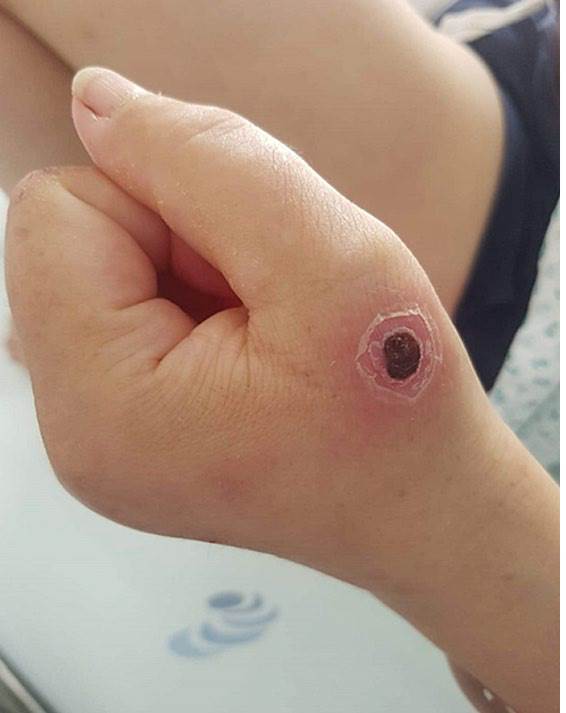
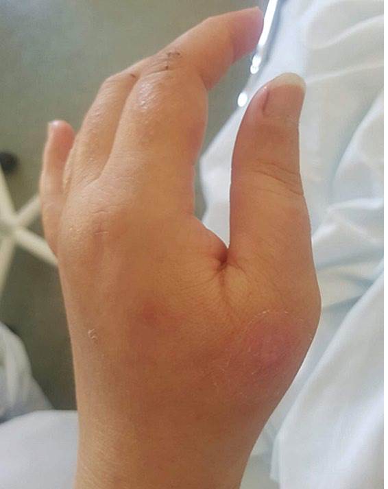
Two days later, due to progressive worsening, she was observed by dermatology. She had several non-painful ulcers with a central black eschar surrounded by an erythematous halo (Fig.s 1A and 1B). The antibiotic was switched to piperacillin/tazobactam (4.5 g i.v. every 6 hours;) with rapid clinical and analytical improvement and complete healing in 7 days (Fig.s 2A and 2B).
Skin and sputum cultures were positive for Pseudomonas aeruginosa, but blood and urine cultures were negative.
Further laboratory evaluation, namely, serological markers to hepatitis B and C, HIV, syphilis, cytomegalovirus, Epstein-Barr virus and parvovirus B19, protein electrophoresis, flow cytometry and bone marrow biopsy were negative.
WHAT IS YOUR DIAGNOSIS?
ECTHYMA GANGRENOSUM
Ecthyma gangrenosum (EG) is an uncommon cutaneous infection, most often seen in immunocompromised patients.Classically, EG is caused by Pseudomonas aeruginosa, an opportunistic agent and one of the leading causes of nosocomial infections, but other bacterial and fungal agents have also been described.1,2
Cutaneous lesions commonly begin as a painless red macule that evolves to a papule, then to an haemorrhagic bullae that ruptures, forming a gray-black eschar with an erythematous halo. The most affected sites are axilla, extremities, gluteal and anogenital areas. The face is rarely affected.1,2
Two clinical forms are described: septic, with hematogenic spread from colonized gastrointestinal, genitourinary or respiratory tracts, and localized, that can result from direct inoculation of the skin or undetected low-grade transient bacteraemia.
Multiple lesions suggest hematogenous dissemination, whereas solitary lesions usually result from direct skin inoculation.1,2
Our patient had a recent diagnosis of FS, a rare, severe extra- articular manifestation of RA, characterized by neutropenia and splenomegaly.1,3Although blood cultures were negative, in our case the presence of multiple disperse lesions suggests a transient bacteraemia.2,5Respiratory symptoms and isolation of Pseudomonas in sputum suggests a respiratory origin. In this case, an upper respiratory infection was initially assumed because of the mild symptoms and normal X-ray, however isolation of Pseudomonas may suggest a lower infection.4 Although no biopsy was performed, the characteristic lesions and the excellent response to treatment support our diagnosis.5
Our case illustrates an atypical presentation of ecthyma gangrenosum, with multiple disperse lesions, including the face, without confirmed bacteraemia. Also noteworthy was the isolation of Pseudomonas aeruginosa in the sputum without radiologic evidence of pneumonia. This highlights the importance of the evaluation of cutaneous lesions, as they were the main key to the correct diagnosis and appropriate treatment.
FS is associated with several dermatological manifestations, however, to our knowledge, there are no reports of EG in these patients.

