SciELO - Scientific Electronic Library Online

 
vol.79 número1Condilomas Acuminados na InfânciaColoração Chicago Sky Blue: Um Novo Aliado no Exame Micológico Directo índice de autoresíndice de assuntosPesquisa de artigos
Home Pagelista alfabética de periódicos  

Serviços Personalizados

Journal

Artigo

Indicadores

Links relacionados

  • Não possue artigos similaresSimilares em SciELO

Compartilhar


Revista da Sociedade Portuguesa de Dermatologia e Venereologia

versão impressa ISSN 2182-2395versão On-line ISSN 2182-2409

Rev Soc Port Dermatol Venereol vol.79 no.1 Lisboa mar. 2021  Epub 15-Jun-2021

https://doi.org/10.29021/spdv.79.1.1267 

Quiz in Dermatology

Pigmented Lesions of the Trunk

Lesões Pigmentadas do Tronco

Joana Barbosa1 
http://orcid.org/0000-0001-7351-5302

Miguel Coelho1 
http://orcid.org/0000-0002-2288-7306

Alexandre João1 

Ana Rodrigues1 

1Dermatology and Venereology Department, Hospital de Santo António dos Capuchos, Centro Hospitalar Universitário de Lisboa Central, Lisbon, Portugal


Introduction

We report the case of a 71-year-old woman diagnosed in the past with a left infiltrating ductal breast carcinoma for which she underwent breast-conserving surgery, axillary lymph node dissection and adjuvant chemotherapy. Thirteen years after surgery, she noticed a pigmented papule which rapidly grew into a hyperkeratotic black plaque surrounding her left nipple. Multiple adjacent black macules were also noticed.

Histopathological analysis revealed intraepidermal and dermal infiltration by atypical epithelioid cells, displaying pagetoid intraepidermal spread. Immunostaining showed positivity for estrogen, progesterone receptors and human epidermal growth factor receptor-type 2 (HER-2). It was also pan-cytokeratin (pan-CK) positive and cytokeratin-7 (CK7) negative.

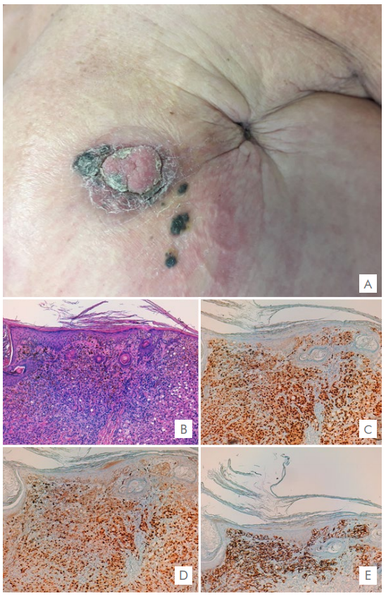
Figure 1 (A) Hyperkeratotic pigmented plaque surrounding the nipple and multiple black macules in the adjacent area; sequel retraction from previous breast-conserving surgery. Skin biopsy (hematoxylin-eosin stain and immunohistochemistry x100) shows: (B) Intraepidermal and dermal infiltration by atypical epithelioid cells, displaying pagetoid intraepidermal spread. Dense pigmentation of the superficial dermis and epidermis. (C) Estrogen receptors; (D) Progesterone receptors; (E) Melan-A showing positivity of dendritic melanocytes surrounding tumor cells. 

Pigmented epidermotropic metastases from breast carcinoma

Cutaneous metastases of a primary internal malignancy are relatively uncommon, accounting for 0.7%‐9% of all metastases. 1 They often occur later in the course of the disease and may manifest years after treatment of the primary tumor. They usually represent a sign of poor prognosis. 2

Breast carcinoma is the most frequent malignancy causing cutaneous metastases3 with a wide range of clinical manifestations and histological appearances. 2 Nodules are the most common clinical presentation but numerous other patterns are described.

Pigmented epidermotropic metastases from breast carcinoma are rare and have not often been described on the literature. 4 They are generally found on the chest and abdominal wall, near the mastectomy scar. 2,5The presence of pigment can be explained by the melanin release from damaged epidermis due to invasion and destruction by tumor cells and consequent phagocytosis by melanophages. 5 Other theories suggest that there is a transfer of melanin from the melanocytes into the carcinoma cells or even that the tumor cells themselves can produce melanin. 2,6,7

On histology, cords, nests and large aggregations of neoplastic cells are present in the dermis and often communicate with the epidermis in a pagetoid spread. 5 The intraepidermal and dermal tumor cells contain melanin in their cytoplasm, and numerous dermal melanophages can be also seen. 4,8

Immunohistochemistry is another helpful tool used to differentiate these findings from clinically indistinguishable entities such as malignant melanoma.8Breast carcinomas are typically cytokeratin 7 positive and cytokeratin 20 negative. 9 S100, HMB45, and Melan-A staining are negative in the majority of cases, in contrast to malignant melanoma. In this case, Melan-A expression was limited to dendritic melanocytes surrounding tumor cells, which did not show cytological atypia or mitotic activity. The presence of estrogen and progesterone hormone receptors and HER2 expression can be evaluated and, as in our case, are frequently positive. 8

In conclusion, this case describes the occurrence of epidermotropic pigmented metastasis manifesting 13 years after the diagnosis of a ductal breast carcinoma. Due to their clinical and histological similarity, pigmented epidermotropic metastases can mimic malignant melanoma which can ultimately lead to a delay in diagnosis of anactive or recurrent internal malignancy. 2 Therefore, the onset of a new pigmented lesion especially close to the surgical scar in patients with an history of internal malignancy should raise suspicion for cutaneous metastasis. Prompt and adequate diagnosis of this condition can significantly impact the patient’s overall prognosis.

References

1. Chraiet N, Zenzri Y, Bouaziz H, Sassi I, Guebsi A, Kamoun S, et al. Generalized cutaneous metastases of breast cancer: An uncommon presentation. Clin Case Rep. 2020;8:667-71. [ Links ]

2. De Giorgi V, Grazzini M, Alfaioli B, Savarese I, Corciova SA, Guerriero G, et al. Cutaneous manifestations of breast carcinoma. Dermatol Ther. 2010;23:581-9. doi: 10.1111/j.1529-8019.2010.01365.x. [ Links ]

3. Wong CYB, Helm MA, Helm TN, Zeitouni N. Patterns of skin metastases: a review of 25 years' experience at a single cancer center. Int J Dermatol. 2014;53:56-60. [ Links ]

4. Requena L, Sangueza M, Sangueza OP, Kutzner H. Pigmented mammary Paget disease and pigmented epidermotropic metastases from breast carcinoma. Am J Dermatopathol.2002;24:189-98. [ Links ]

5. Brasanac D, Boricic I, Todorovic V. Epidermotropic metastases from breast carcinoma howing different clinical and histopathological features on the trunk and on the scalp in a single patient. J Cutan Pathol. 2003;30:641-6. doi: 10.1034/j.1600-0560.2003.00130.x. [ Links ]

6. Hamada M, Toyoshima S, Duan H, Furue M. Pigmented cutaneous metastasis of mucinous carcinoma of the breast to the scalp mimicking malignant melanoma. Eur J Dermatol.2006;16:592-3. [ Links ]

7. Ishihara-Yusa S, Fujimura T, Lyu C, Sugawara M, Sakamoto K, Aiba S. Breast cancer metastasis in the skin with hyperkeratotic pigmentation caused by melanocyte colonization. Case Rep Oncol. 2018;11:660-4. doi: 10.1159/000493186. [ Links ]

8. Ubillos N, Vola M, Mazzei ME, Magliano J. Pigmented Cutaneous Metastasis of Breast Carcinoma Mimicking a Melanoma. Actas Dermosifiliogr. 2016;107:699-701. doi: 10.1016/j.ad.2016.04.007. [ Links ]

9. Lee AHS. The histological diagnosis of metastases to the breast from extramammary malignancies. J Clin Pathol. 2007;60:1333-41. [ Links ]

Responsabilidades ÉticasConflitos de Interesse: Os autores declaram a inexistência de conflitos de interesse na realização do presente trabalho. Fontes de Financiamento Suporte Financeiro: Não existiram fontes externas de financiamento para a realização deste artigo. Confidencialidade dos Dados: Os autores declaram ter seguido os protocolos da sua instituição acerca da publicação dos dados de doentes. Proteção de Pessoas e Animais: Os autores declaram que os procedimentos seguidos estavam de acordo com os regulamentos estabelecidos pelos responsáveis da Comissão de Investigação Clínica e Ética e de acordo com a Declaração de Helsínquia da Associação Médica Mundial. Proveniência e Revisão por Pares: Não comissionado; revisão externa por pares

Ethical DisclosuresConflicts of Iinterest: The authors have no conflicts of interest to declare. Financing Support: This work has not received any contribution, grant or scholarship Confidentiality of Data: The authors declare that they have followed the protocols of their work center on the publication of data from patients. Protection of Human and Animal Subjects: The authors declare that the procedures followed were in accordance with the regulations of the relevant clinical research ethics committee and with those of the Code of Ethics of the World Medical Association (Declaration of Helsinki). Provenance and Peer Review: Not commissioned; externally peer reviewed.

3© Author(s) (or their employer(s)) and SPDV Journal 2020. Re-use permitted under CC BY-NC. No commercial re-use. © Autor (es) (ou seu (s) empregador (es)) e SPDV Revista 2020. Reutilização permitida de acordo com CC BY-NC. Nenhuma reutilização comercial

Received: August 24, 2020; Accepted: October 03, 2020

Autor Correspondente/Corresponding Author: Joana Barbosa E-mail: joana.barbosa@chlc.min-saude.pt Address: Serviço de Dermatologia do Hospital de Santo António dos Capuchos, Alameda Santo António dos Capuchos, 1169-050 Lisboa, Portugal

Creative Commons License This is an open-access article distributed under the terms of the Creative Commons Attribution License