


 Inglês (pdf)
 Artigo em XML
 Referências do artigo
 Enviar este artigo por email
 Citado por SciELO
  Similares em
SciELO
Similares em
SciELO 
 Permalink
INTRODUCTION
Schwannoma, also known as neurilemmoma, is a benign encapsulated neoplasm composed of Schwann cells that originates from nerve sheaths.1 Commonly found in association with the cranial nerves (especially the acoustic nerve), schwannomas may arise anywhere along the course of a nerve, including peripheral nerves.2Cutaneous schwannoma usually presents as an asymptomatic well-circumscribed skin-colored nodule of slow growth, located in the dermis or in the subcutaneous tissue.1,2The most common sites are the head and neck and the flexor surfaces of the extremities.3,4Although they commonly occur as single lesions (90%), they can be associated with several central neurological tumors (usually meningiomas, 5%), neurofibromatosis type 2 (3%), or appear as multiple lesions (schwannomatosis, 2%).3
Herein, we present a case of a solitary cutaneous ancient schwannoma of the ear, a histopathological variant of schwannoma with distinctive morphological characteristics.
CASE REPORT
An otherwise healthy 69-year-old caucasian woman was observed for a painless skin nodule on her left ear, that had developed slowly over a period of more than eight years. The patient denied previous local trauma or any other similar lesions. Physical examination revealed a well-defined, flesh-colored, cutaneous nodule, 1 cm in diameter, with elastic consistency, non-painful on palpation, located on the patient’s concha of the left ear (Fig. 1). There were no other relevant skin lesions.
Surgical excision with clear margins was performed. Histological examination revealed an encapsulated tumor located in the upper dermis composed of mostly dense cellular areas (Antoni type A), alternating with hypocellular myxoid areas (Antoni type B) (Fig. 2 A,B). On high-power magnification, the tumor was composed of spindle cells with back to back nuclei arranged in parallel rows without mitoses. There were some enlarged, hyperchromatic, and bizarre nuclei (Fig. 2C).
Conspicuous small blood vessels were seen. Immunohistochemically, the tumor cells were strongly and diffusely immunorreactive for S-100 protein (Fig. 2D), and the tumor capsule cells stained for EMA (Fig. 2E).
Alcian blue showed staining of the myxoid matrix (Fig. 2F). Based on histological examination, a diagnosis of ancient schwannoma was made.
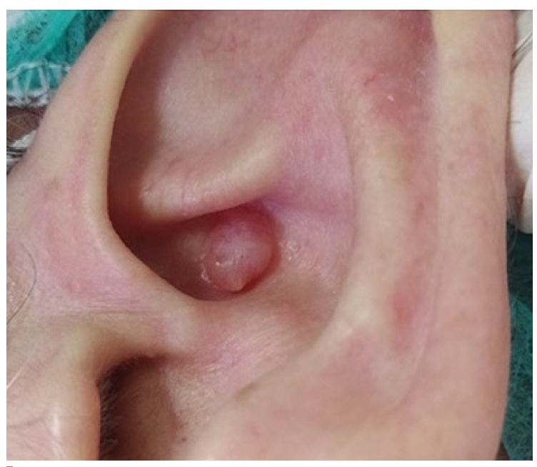
Figure 1 Cutaneous schwannoma, clinical picture: Cutaneous nodule with 1cm in diameter on the patients’ left concha of the ear.
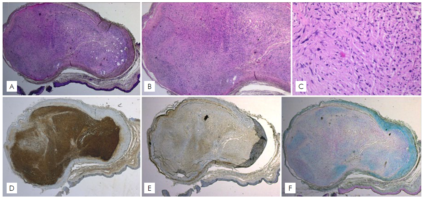
Figure 2 Cutaneous ancient schwannoma. The upper dermis showed a biphasic encapsulated intradermal tumor (A. H&E x16) with mostly hypercellular zones (Antoni A pattern) and with some hypocellular zones (Antoni B pattern) (B. H&E x40). The tumor was composed of spindle cells, some with enlarged, hyperchromatic, and bizarre nuclei but no mitotic activity (C. H&E x100). Cells showed positivity for S100 (D. S100 x16) and fibroblasts in the perineurium sheath were positive for EMA (E. EMA x16). The myxoid stroma stained for Alcian blue (F. Alcian blue x16).
DISCUSSION
Verocay first described schwannomas in 1908.3 They represent approximately 5% of all benign soft-tissue tumors and they are the most common peripheral nerve tumor.4 Schwannomas occur most commonly during the fourth and fifth decade of life, without significant evidence of sex predilection.3-5
In general, cutaneous schwannomas are asymptomatic.5 However, as they grow larger, pain, paresthesias, and tenderness can occur, associated with the compression of the nerve of origin or adjacent structures.5
The clinical diagnosis is challenging due to the non-specific presentation, becoming necessary to exclude other well-circumscribed skin masses such as lipoma, angiolipomas, leiomyomas, cysts, neurofibromas, and neuromas.3
Histology is required to confirm the diagnosis. The tumor is typically well encapsulated by perineurium and is classically characterized by a biphasic pattern: areas of high cellularity (Antoni A) that alternate with loosely myxoid edematous areas (Antoni B) (Fig. 3).1-6 In Antoni A areas, uniform spindle cells are clustered in stacks and arranged back to back, creating palisades of stacked nuclei between which the cell processes are fused into eosinophilic masses, called Verocay bodies (Fig. 3 A, B).1-6In Antoni B areas, cells are more loosely arranged, separated by spaces filled with mucin. (Fig. 3 C, D).1-6Most schwannomas display both areas, and although intermingled, both region types usually appear fairly demarcated from one another.5
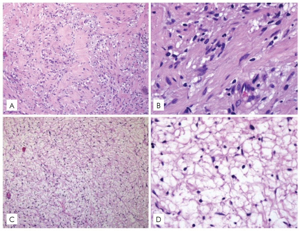
Figure 3 A,B. Antoni A area: dense spindle cells arranged in an orderly fashion. The nuclei are aligned in palisades, forming the distinctive Verocay bodies. (A. H&E, x100; B. H&E, x400); C,D. Antoni B area: scattered spindle cells in loose and myxoid stroma. (C. H&E x100, D. H&E x400).
The immunohistochemical profiles of schwannomas are typically positive for S100 protein and collagen type IV, while the fibroblast in the perineurium sheath is positive for epithelial membrane antigen (EMA).1,6As most of schwannomas are devoid of axons, they are characteristically negative for neurofilaments.1
Ancient schwannoma is a rare subtype of schwannoma, which is usually found in long-standing lesions, characterized by pronounced degenerative changes.7-10In addition to the classical features exhibited in schwannoma, ancient type displays the presence of enlarged, hyperchromatic and bizarre nuclei and the absence of mitosis.7-10
Cystic formation, calcification, haemorrhage and hyalinization may also be demonstrated.7-10The characteristic histologic appearance seen in ancient schwannoma can make pathologic diagnosis difficult and lead to an erroneous diagnosis of malignancy.7-10
Although ancient schwannomas are a well-known entity that can involve the head and neck region, with reports in a variety of locations, such as the orbit, infratemporal fossa, submandibular gland and parotid gland,10 but it is extremely rare for them to be found in the skin of of the ear.
The treatment of choice for cutaneous schwannoma is surgical excision, indicated in the presence of symptoms or for cosmetic reasons.5 Local recurrence is infrequent as the tumor is usually well-encapsulated and thus easily resectable.5 Malignant transformation of solitary schwannomas is exceedingly rare.6 The prognosis of ancient schwannoma is similar to the classic variant so atypia should not raise alarm for malignant transformation.8
In conclusion, cutaneous schwannomas are benign skin tumors with a very characteristic histology but non-specific clinical presentation. We highlight the importance of recognizing the histopathological variant of ancient schwannoma to avoid an erroneous diagnosis of malignancy and unnecessarily aggressive treatment.

