SciELO - Scientific Electronic Library Online

 
vol.79 número2Lesão Hiperqueratósica Plantar Dolorosa índice de autoresíndice de assuntosPesquisa de artigos
Home Pagelista alfabética de periódicos  

Serviços Personalizados

Journal

Artigo

Indicadores

Links relacionados

  • Não possue artigos similaresSimilares em SciELO

Compartilhar


Revista da Sociedade Portuguesa de Dermatologia e Venereologia

versão impressa ISSN 2182-2395versão On-line ISSN 2182-2409

Rev Soc Port Dermatol Venereol vol.79 no.2 Lisboa jun. 2021  Epub 01-Out-2021

https://doi.org/10.29021/spdv.79.2.1378 

Letter to the Editor

Furuncular Myiasis of the Scalp, an Unexpected Diagnosis

Miíase Cutânea Furuncular do Couro Cabeludo, um Diagnóstico Inesperado

1Serviço de Pediatria, Centro Hospitalar Barreiro-Montijo, Barreiro, Portugal


We report the case of 10-year-old Caucasian child with no relevant personal history living in a farm with basic sanitation conditions, where pigs were bred. She reported a nodule on her right occipital region that, within 24-48 hours, turned erythematous with a small opening on the top in association with multiple adenopathies in the right cervical region. Oral amoxicillin/clavulanic acid and topical fusidic acid were prescribed, but within the edematous furunculoid nodule the central opening enlarged, and two larvae could be seen moving inside the hole (Fig. 1). The diagnosis of furuncular myiasis was considered and mechanical extraction of larvae was performed, leaving the cavity apparently clean. An oral dose of ivermectin (150 μg/kg) was prescribed and one month later, the wound was completely healed.

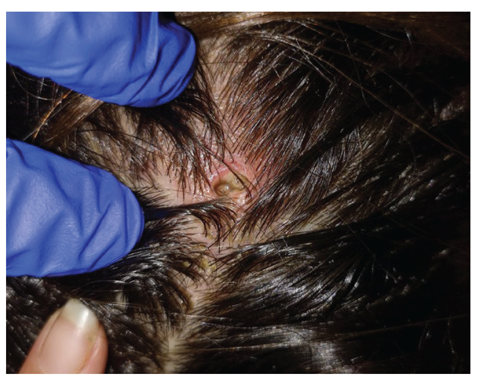
Figure 1 Inflammatory nodule on the scalp with a central ulcer where two larvae could be seen moving. 

Myiasis is an infestation of the skin and mucous membranes by dipterous maggots, namely Dermatobia hominis, the human botfly, which can produce furuncular (the most common), creeping, or traumatic lesions.1-5Human myiasis is a worldwide infection, mostly related to warm and humid environment and frequently related to previous travel history, but other risk factors including poor hygiene and low socioeconomic status may favour the disease,3-5as in this case.

Lesions are normally single and usually harbour only one larva, with the breathing tube visible within the skin hole. Lesions occur most commonly on exposed areas, so scalp lesions are uncommon.3-5

Myiasis' diagnosis is clinical, but dermoscopy can be helpful.3,5

Treatment consists on the mechanic removal of the larva usually after application of a toxic substance.4,5 If larvae are not accessible, skin occlusion may be necessary to promote localized hypoxia and consequent larval migration to skin surface.4,5Surgical excision may be needed when dead larvae remain inside the skin. Oral treatment of human myiasis is not consensual, but ivermectin (single oral dose 150-200 μg/kg) is the main choice.6

This case of furuncular myiasis highlights some atypical findings, particularly the location on the scalp, and the rural and poor socioeconomic condition as another risk factor in the absence of previous travelling.

REFERENCES

1. Lachish T, Marhoom E, Mumcuoglu KY, Tandlich M, Schwarts E. Myiasis in travelers. J Travel Med. 2015;22:232-6. doi:10.1111/jtm.12203. [ Links ]

2. Mammino J, Lal K. Myiasis: A Traveler's Dilemma. J Clin Aesthet Dermatol. 2013;6:47-9. [ Links ]

3. Francesconi F, Lupi O. Myiasis. Clin Microbiol Rev. 2012;25:79-105. doi:10.1128/CMR.00010-11. [ Links ]

4. Palmieri JR, North D, Santo A. Furuncular myiasis of the foot caused by the tumbu fly, Cordylobia anthropophaga report in a medical student returning from a medical mission trip to Tanzania. Int Med Case Rep J. 2013;6:25-8. doi: 10.2147/IMCRJ.S44862. [ Links ]

5. How EH, Yap D, Mbakada N. An exotic abscess within the United Kingdom from the Gambia: A case report. J Med Case Rep. 2017;11:310. doi: 10.1186/s13256-017-1472-3. [ Links ]

6. Cunha PR, Flora TB, Kroumpouzos G. Travelers' tropical skin diseases: Challenges and interventions. Dermatol Ther. 2019;32:e12665. doi: 10.1111/dth.12665 [ Links ]

1© Author(s) (or their employer(s)) 2021 SPDV Journal. Re-use permitted under CC BY-NC. No commercial re-use. © Autor (es) (ou seu (s) empregador (es)) 2021 Revista SPDV. Reutilização permitida de acordo com CC BY-NC. Nenhuma reutilização comercial

2Conflicts of Interest: The authors have no conflicts of interest to declare. Financing Support: This work has not received any contribution, grant or scholarship. Confidentiality of Data: The authors declare that they have followed the protocols of their work center on the publication of data from patients. Patient Consent: Consent for publication was obtained. Provenance and Peer Review: Not commissioned; externally peer reviewed. Conflitos de Interesse: Os autores declaram a inexistência de conflitos de interesse na realização do presente trabalho. Suporte Financeiro: Não existiram fontes externas de financiamento para a realização deste artigo. Confidencialidade dos Dados: Os autores declaram ter seguido os protocolos da sua instituição acerca da publicação dos dados de doentes. Consentimento: Consentimento do doente para publicação obtido. Proveniência e Revisão por Pares: Não comissionado; revisão externa por pares

Received: March 09, 2021; Accepted: March 16, 2021

Corresponding Author: Gonçalo Vale Address: Rua Dr. Ramiro Barros Lima, lote 3, 4740-267, Esposende - Portugal E-mail:gnpvale14@gmail.com

Creative Commons License This is an open-access article distributed under the terms of the Creative Commons Attribution License