SciELO - Scientific Electronic Library Online

 
vol.32 issue1Bone Scintigraphy in McCune-Albright SyndromeEctopic Intrathyroidal Thymus Mimicking a Thyroid Nodule author indexsubject indexarticles search
Home Pagealphabetic serial listing  

Services on Demand

Journal

Article

Indicators

Related links

  • Have no similar articlesSimilars in SciELO

Share


Acta Radiológica Portuguesa

Print version ISSN 2183-1351

Acta Radiol Port vol.32 no.1 Lisboa Apr. 2020  Epub June 30, 2021

https://doi.org/10.25748/arp.18777 

Imagens de Interesse

Post-Biopsy Arteriovenous Fistula in Kidney Transplant

Fístula Arteriovenosa Pos-Biópsia em Rim Transplantado

1Department of Radiology, Centro Hospitalar Universitário Lisboa Central, Hospital Curry Cabral, Lisboa, Portugal


Abstract

A 53-year-old patient submitted to a kidney transplant 5 years ago presented with hematuria after kidney biopsy for transplant dysfunction. Color-Doppler ultrasound was performed and revealed an arteriovenous fistula in the kidney transplant parenchyma.

Keywords: Renal arteriovenous fistula; Kidney transplant; Color doppler ultrasound

Resumo

Uma mulher de 53 anos, submetida a transplante renal 5 anos antes, apresenta hematúria após realizar biópsia do transplante por disfunção do enxerto. O eco-Doppler a cores revelou a presença de uma fístula arteriovenosa no parênquima do enxerto renal.

Palavras-chave: Fístula arteriovenosa renal; Transplante renal; Eco-doppler a cores.

A 53-year-old patient submitted to a renopancreatic transplant 5 years ago presented progressive asymptomatic renal transplant dysfunction (Creatinine 1.9 → 2.6mg/dL) and increased arterial resistive index (0,88). A transplantbiopsy was performed revealing cellular rejection type IIA and thymoglobulin was initiated. After biopsy the patient presented macroscopic hematuria and Color-Doppler ultrasound revealed an arteriovenous fistula (AVF) in the kidney transplant parenchyma.

Embolization was postponed due to fever and E. Faecalis isolation on hemocultures, treated with ampicillin, with clinical and laboratorial improvement. A new biopsy was performed and showed no signs of cellular rejection. Serum antibodies were also negative for alloimmunization(Figs. 1, 2).

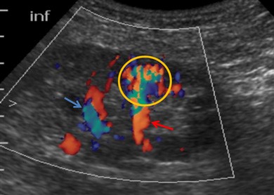
Figure 1: Color Doppler ultrasou- nd depicting an AVF (yellow circle) with aliasing due to turbulent flow and its arterial and venous vessels (red and blue arrows, respectively). 

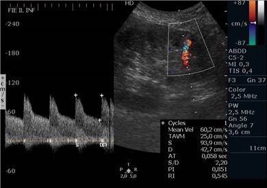
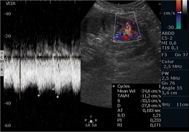
Figure 2: Color and Spectral Doppler from the artery feeding the fistula showing high peak systolic velocity (94cm/s) and a low¬-resistance pattern with increased diastolic velocity (42cm/s). 

The patient remained clinically stable after 1 year of biopsy and showed progressive improvement of renal function, despite persistence of a high-debit fistula, therefore embolization was not performed and the patient remains in follow up.

Core needle biopsy remains an important tool in evaluation of renal transplant dysfunction.1 AVF is a common complication following biopsy or percutaneous vascular procedures.1,2 AVF can be asymptomatic or manifest as hematuria, renal failure or hypertension.3 On Color -Doppler, AVF appears as a focal high-velocity flow showing aliasing, that communicates with a feeding artery, with accentuated diastolic flow and a draining vein, with an arterialized venous waveform.2,3 Most small AVFs resolve spontaneously and only larger and symptomatic AVFs may need transcatheter embolization(Fig. 3).1,3

Figure 3: Color and Spectral Doppler form the vein draining the fistula showing an arterialized flow. 

References

1. Schwarz A et al. Course and relevance of arteriovenous fistulas after renal transplant biopsies. American Journal of Transplantation. 2008;8: 826-31. [ Links ]

2. Kaufman JA, Lee MJ. Vascular & interventional radiology: The Requisites. Saunders Inc. 2014. [ Links ]

3. LaBerge JM. Interventional management of renal transplant arteriovenous fistula. Seminars in Interventional Radiology. 2004;21:239- 46. [ Links ]

Ethical disclosures

Financing Support: This work has not received any contribution, grant or scholarship.

Received: October 26, 2019; Accepted: December 20, 2019

Address Ana Luísa Proença Serviço de Radiologia, Centro Hospitalar Universitário Lisboa Central, Hospital Curry Cabral, Rua Beneficência 8, 1050-099 Lisboa, Portugal email: proenca.ana.luisa@gmail.com

Conflicts of interest: The authors have no conflicts of interest to declare.

Confidentiality of data: The authors declare that they have followed the protocols of their work center on the publication of data from patients.

Protection of human and animal subjects: The authors declare that the procedures followed were in accordance with the regulations of the relevant clinical research ethics committee and with those of the Code of Ethics of the World Medical Association (Declaration of Helsinki).

Creative Commons License This is an open-access article distributed under the terms of the Creative Commons Attribution License