SciELO - Scientific Electronic Library Online

 
vol.32 issue1Post-Biopsy Arteriovenous Fistula in Kidney Transplant author indexsubject indexarticles search
Home Pagealphabetic serial listing  

Services on Demand

Journal

Article

Indicators

Related links

  • Have no similar articlesSimilars in SciELO

Share


Acta Radiológica Portuguesa

Print version ISSN 2183-1351

Acta Radiol Port vol.32 no.1 Lisboa Apr. 2020  Epub June 30, 2021

https://doi.org/10.25748/arp.19163 

Imagens de Interesse

Ectopic Intrathyroidal Thymus Mimicking a Thyroid Nodule

Timo Intratiroideu Ectópico que Mimetiza um Nódulo da Tiroide

Daniela Barros1 

Ana Catarina Costa1 

Fernanda Marta Gomes1 

Catarina Costa1 

Vasco Mendes1 

1Department of Radiology, Hospital de Braga, Braga, Portugal


Abstract

Ectopic thymus is a well-known embryological abnormality in children, despite being considered rare. To avoid unnecessary examination or biopsy, radiologists must be aware of this condition, especially in pediatric population. This report describes a pediatric patient with history of recurrent episodes of tonsillitis since 2 years of age. In the context of infectious mononucleosis, a cervical ultrasound was performed, which revealed a relatively hypoechoic lesion in the right lobe of the thyroid gland, similar to normal thymus echogenicity. Thyroid function tests were normal. After 3 years of follow-up, the intrathyroidal nodule maintained the same characteristics, having decreased in size from 12 mm to 8 mm.

Keywords: Ectopic thymus; Thymus; Thyroid; Ultrasound.

Resumo

O timo ectópico é uma anormalidade embriológica bem conhecida em crianças, apesar de ser considerado raro. Para evitar biópsias desnecessárias, os radiologistas devem estar cientes dessa condição, principalmente na população pediátrica. Este artigo descreve um paciente pediátrico com história de episódios recorrentes de amigdalite desde os 2 anos de idade. No contexto de uma mononucleose infeciosa foi realizada uma ecografia cervical, que revelou uma lesão relativamente hipoecóica no lobo direito da glândula tireóidea, semelhante à ecogenicidade normal do timo. Os testes de função tireóidea foram normais. Após 3 anos de acompanhamento, o nódulo intratiroideu mantém as características iniciais e diminuiu de tamanho de 12 mm para 8 mm.

Palavras-chave: Ecografia; Timo ectópico; Timo; Tiroide.

Introduction

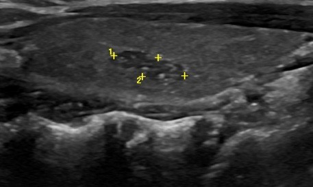
We present a case of a pediatric patient with history of recurrent episodes of tonsillitis since 2 years of age. In the context of infectious mononucleosis, a cervical ultrasound was performed, which revealed a relatively hypoechoic lesion in the right lobe of the thyroid gland, similar to normal thymus echogenicity (Fig. 1). Thyroid function tests: TT3 (total triiodothyronine), TT4 (total tetraiodothyronine), FT3 (free triiodothyronine), FT4 (free tetraiodothyronine) and TSH (thyroid-stimulating hormone) were normal. After 3 years of follow-up, the intrathyroidal nodule maintained the same characteristics, having decreased in size from 12 mm to 8 mm (Fig. 2). Decision of conservative management with strict monitoring until involution was taken, along with parents’ consent.

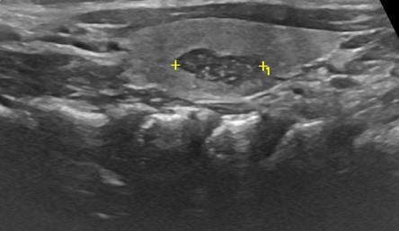
Figure 1: Ultrasound showing a relatively hypoechoic 12mm lesion in the right lobe of the thyroid gland, with sharp margins and multiple echogenic foci, similar to thymus tissue. 

Figure 2: Ultrasound after 3 year follow up, showing the intra- thyroidal nodule with the same characteristics, having decreased in size, from 12 mm to 8 mm. 

Role of ultrasound in diagnosis and follow up of the ectopic thymus

As ultrasound is of common use nowadays, the number of incidental lesions found is increasingly high, including simple cysts or hypoechoic nodules, such as ectopic thymus. Ectopic thymus is a well-known embryological abnormality in children, despite being considered rare. It is most frequently found in the cervical area or mediastinum, yet rarely in the pericardium, hilar area, chest wall or axilla. Ectopic intrathyroidal thymic tissue is also rare.1

Normal thymus tissue, located in the superior and anterior compartments of the mediastinum, develops from the endoderm of the third and fourth pharyngeal pouches. Thymic parenchyma in children has a characteristic ultrasound appearance, with fusiform shape, well-defined margins and echogenicity between the thyroid gland and the surrounding muscle. Furthermore, it has multiple echogenic foci, portraying a “starry sky” appearance. This distinctive feature is created by overlapped fat and lymphoid tissue.2 So, if an intrathyroidal lesion has sharp margins and echotexture similar to the thymus, with no abnormal echogenicities in the surrounding thyroid area,as well as no abnormal lymph nodes, it requires no further action. Only sonographic follow-up is advised, since thymic tissue may automatically degrade and contract. However, if the diagnosis is uncertain, based on sonographic features, or if the mass has suspicious characteristics, further investigation, including ultrasound-guided fine-needle aspiration or additional imaging should be performed.3

In conclusion, it is important to identify and to be aware of thymic rests and their normal sonographic appearance, so that they won’t be confused with other neoplastic thyroid masses, therefore avoiding unnecessary anxiety and intervention, especially in the pediatric population.

References

1. Huang Y, Zheng S, Xiao X. Ectopic intrathyroidal thymus in children: two case reports and review of the literature. J Ped Surg Case Reports. 2013;1:386-90. [ Links ]

2. Kim HG, Kim MJ, Lee MJ. Sonographic appearance of intrathyroid ectopic thymus in children. J Clin Ultrasound. 2012;40:266-71. [ Links ]

3. Megremis S, Stiakaki E, Tritou I, et al. Ectopic intrathyroidal thymus misdiagnosed as a thyroid nodule: sonographic appearance. J Clin Ultra- sound. 2008;36:443-7. [ Links ]

Ethical disclosures

Financing Support: This work has not received any contribution, grant or scholarship.

Received: December 31, 2019; Accepted: January 10, 2020

Address Daniela Barros Serviço de Radiologia Hospital de Braga Lugar das Sete Fontes, 4710-243 Braga, Portugal email: danielabarros93@gmail.com

Conflicts of interest: The authors have no conflicts of interest to declare.

Confidentiality of data: The authors declare that they have followed the protocols of their work center on the publication of data from patients.

Protection of human and animal subjects: The authors declare that the procedures followed were in accordance with the regulations of the relevant clinical research ethics committee and with those of the Code of Ethics of the World Medical Association (Declaration of Helsinki).

Creative Commons License This is an open-access article distributed under the terms of the Creative Commons Attribution License