SciELO - Scientific Electronic Library Online

 
vol.32 issue2Fibrodysplasia Ossificans Progressiva: Stone Man Syndrome author indexsubject indexarticles search
Home Pagealphabetic serial listing  

Services on Demand

Journal

Article

Indicators

Related links

  • Have no similar articlesSimilars in SciELO

Share


Acta Radiológica Portuguesa

Print version ISSN 2183-1351

Acta Radiol Port vol.32 no.2 Lisboa Aug. 2020  Epub July 08, 2021

https://doi.org/10.25748/arp.19827 

Imagens de Interesse

OHVIRA Syndrome with a Blind-Ended Ureteral Remnant

Síndrome OHVIRA com Remanescente Uretérico

1Radiology Department, Hospital do Divino Espírito Santo de Ponta Delgada, EPER, Ponta Delgada, Portugal

2Radiology Department, Hospital Dona Estefânia, Centro Hospitalar de Lisboa Central, EPE, Lisboa, Portugal

3Head of Service. Radiology Department, Hospital Dona Estefânia, Centro Hospitalar de Lisboa Central, EPE, Lisboa, Portugal


Abstract

OHVIRA syndrome is characterized by a didelphys uterus with an obstructed/blind hemi-vagina and ipsilateral renal agenesis. We presented a case of a female child with a pre- natal diagnosis of left renal agenesis whose post-natal imaging findings were consistent with OHVIRA syndrome.

Keywords: OHVIRA syndrome; Mullerian malformations; Congenital female genital tract anomalies.

Resumo

O Síndrome OHVIRA é caracterizado pela presença de útero didelfos com uma hemi- vagina obstruída e agenesia renal ipsilateral. Apresentamos o caso de uma criança com diagnóstico pré-natal de agenesia renal esquerda cuja investigação imagiológica pós-natal revelou um Síndrome OHVIRA.

Palavras-chave: Síndrome OHVIRA; Malformações mullerianas; Anomalias congénitas genitais femininas.

Ten-day-old female referred to a pediatric urology consultation due to prenatal diagnosis of left renal agenesis and a pelvic cystic image, which raised the suspicion of a nephro-urologic malformation.

At birth, analytically with normal values of Urea (34 mg/dl) and Creatinine (1.38 mg/dl). Ultrasound in the first week of life confirmed a hydrohematocolpos, showing a pelvic, non-pure, cystic image (Fig. 1), identified two hemi-uterus suggesting a didelphys uterus and a tubular image with bladder insertion adjacent to the left uterus, interpreted as a ureteral remnant.

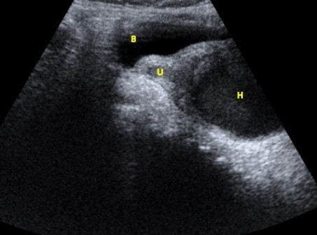
Figure 1: Pelvic ultrasound (longitudinal) showing the blader (B), the left hemi-uterus (U) and a hematocolpos of heterogenous content in the obstructed hemi-vagina (H). 

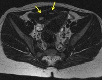
Hydrohematocolpos was drained to prevent infection. Magnetic resonance imaging was performed at 7 months of age, confirming left renal agenesis, a left ureteral remnant with ureterocele (Fig. 2) and a didelphys uterus (Fig. 3).

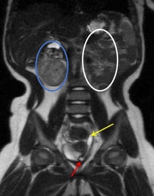
Figure 2: Coronal T2-weighted image demonstrating the presence of a right kidney (blue circle), absence of the left kidney (white circle), a left ureteral remnant (yellow arrow) and an ureterocele (red arrow). 

Figure 3: Axial T2-weighted image showing two hemi-uterus (arrows). 

Currently at 2 years-old, she remains asymptomatic and maintaining regular follow-ups with urologic pediatricians. OHVIRA (Obstructed hemivagina and ipsilateral renal anomaly) syndrome, also known by Herlyn-Werner- Wunderlich syndrome, is a congenital anomaly of the female urogenital tract resulting from an anomaly of the Mullerian and mesonephric ducts. It is characterized by a didelphys uterus with an obstructed/blind hemi-vagina and ipsilateral renal agenesis1 accounting for 0.16-10% of Mullerian duct malformations (MDM).

Patients are usually asymptomatic until puberty, when they begin to have dysmenorrhea and cyclic pelvic pain due to hematometrocolpos, as a consequence of obstructed hemi- vagina. In the postnatal period and early infancy, the same symptoms may occur due to the influence of maternal hormones.

Acute complications include pyohematocolpos and pyosalpinx, and long-term complications include endometriosis, pelvic inflammatory disease, and infertility.1 Differential diagnosis includes others MDM (bicornuate uterus, septate uterus), imperforate hymen and transverse vaginal septum.

Treatment is symptomatic, with vaginal septotomy for hematocolpos drainage. Hemi-hysterectomy is not indicated.2,3

As there is a strong association of female genital tract and renal anomalies, when the patient presents with one of these, a screening for associated anomalies should be performed.

References

1. Del Vescovo R, Battisti S, Di Paola V, Piccolo CL, Cazzato RL, Sansoni I et al. Herlyn-Werner-Wunderlich syndrome: MRI findings, radiological guide (two cases and literature review), and differential diagnosis. BMC Medical Imaging 2012;12:4. [ Links ]

2. Ahmad Z, Goyal A, Das CJ, Deka D, Sharma R. Herlyn-Werner- Wunderlich syndrome presenting with infertility: Role of MRI in diagnosis. Indian Journal of Radiology and Imaging 2013;23:3. [ Links ]

3. Cho YH, Sung DJ, Han NY, Park BJ, Kim MJ, Sim KC et al. MRI findings of obstructed hemivagina and ipsilateral renal agenesis (OHVIRA syndrome) with a blind megaureter: Case report. iMRI 2015;19:196-9. [ Links ]

Ethical disclosures

Financing Support: This work has not received any contribution, grant or scholarship.

Received: April 04, 2020; Accepted: May 12, 2020

Address Rafaela Sousa Serviço de Radiologia, Hospital do Divino Espírito Santo de Ponta Delgada, Avenida D. Manuel I 9500-370 Ponta Delgada, Portugal email: rafaelasousa090@hotmail.com

Conflicts of interest: The authors have no conflicts of interest to declare.

Confidentiality of data: The authors declare that they have followed the protocols of their work center on the publication of data from patients.

Protection of human and animal subjects: The authors declare that the procedures followed were in accordance with the regulations of the relevant clinical research ethics committee and with those of the Code of Ethics of the World Medical Association (Declaration of Helsinki).

Creative Commons License This is an open-access article distributed under the terms of the Creative Commons Attribution License