SciELO - Scientific Electronic Library Online

 
vol.33 número3Variabilidade Sazonal da Captação de [18F]FDG em Gordura Castanha na mesma DoenteNervo Cubital Bipartido - Uma Variação Anatómica Rara índice de autoresíndice de materiabúsqueda de artículos
Home Pagelista alfabética de revistas  

Servicios Personalizados

Revista

Articulo

Indicadores

Links relacionados

  • No hay articulos similaresSimilares en SciELO

Compartir


Acta Radiológica Portuguesa

versión impresa ISSN 2183-1351

Acta Radiol Port vol.33 no.3 Lisboa dic. 2021  Epub 31-Dic-2021

https://doi.org/10.25748/arp.25235 

Imagens de Interesse

Post-Traumatic Thrombosis of Pampiniform Plexus

Trombose Pós-Traumática do Plexo Pampiniforme

Catarina Ala Baraças1 
http://orcid.org/0000-0002-3595-2015

Abel Salgueiro1 

Maria Catarina Tavares1 

1Department of Radiology, Hospital Pedro Hispano, ULS Matosinhos, Matosinhos, Portugal


Abstract

Acute scrotal pain is a frequent cause of admission in the emergency room. However, painful scrotum due to pampiniform plexus thrombosis is rare.

The authors discuss a case of a 44-year-old man who presented to the emergency department with left scrotal pain with 5 days of duration, after falling from his own height in a sitting position.

Keywords: Acute scrotum; Thrombosis; Color doppler ultrasound; Testicular vein.

Resumo

A dor escrotal aguda é uma causa frequente de admissão no serviço de urgência. No entanto, a trombose do plexo pampiniforme é uma causa rara de dor escrotal.

Os autores apresentam o caso de um homem de 44 anos que recorre ao serviço de urgência por dor escrotal esquerda com 5 dias de evolução, após queda da própria altura em posição sentado.

Palavras-chave: Escroto aguda; Trombose; Ecografia doppler; Veia testicular.

Case Presentation

A 44-year-old man presented to the emergency department complaining of left scrotal pain, 5 days after falling from his own height in a sitting position. The patient referred a gradual worsening of the pain, without fever or other symptoms.

Physical examination revealed a little enlargement and tenderness in the left hemiscrotum, with a painful lump in the inferior portion of the testicle, which did not change after Valsalva maneuver.

Laboratory analysis signaled no relevant inflammatory markers.

A scrotal Doppler Ultrasound exam was performed, and when the probe was placed directly over the point of maximal tenderness, as indicated by the patient, it immediately revealed a dilated, noncompressible pampiniform plexus’ vein with echogenic intraluminal filling defect, and absent flow at color Doppler (Figure 1).

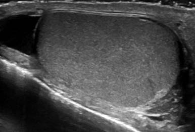
Mild hydrocele was also documented (Figure 2)

Both testicles and epididymis were symmetrical and had normal echotexture.

The patient underwent analgesic and anticoagulation treatment (enoxaparin - low molecular weight heparin) for 2 weeks, with bed rest, with good clinical results.
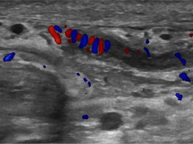
Figure 1: Left spermatic cord with a dilated, noncompressible pampiniform plexus’ vein with echogenic intraluminal filling defect (arrow). 

Figure 2: Mild left hidrocele with normal left testicle 

Discussion

Spontaneous thrombosis of the pampiniform plexus is an uncommon entity responsible for painful scrotum. Left side thrombosis is most common. Anatomical particularities seem to justify left side preponderance.1

Pampiniform plexus thrombosis is being associated with coagulation abnormalities, retroperitoneal tumors, vigorous exercise, or anatomical factors, such as compression of the left renal vein by superior mesenteric artery (nutcracker syndrome), absent or incompetent valves of the testicular vein, or absence of inferior vena cava.2,3Although trauma is included in several articles as a cause of pampiniform plexus thrombosis, the authors did not find any similar case report.

Doppler ultrasound shows a tubular noncompressible structure, with more or less echogenic content, depending on temporal evolution, with no blood flow.

With spontaneous thrombosis of the pampiniform plexus, some differential diagnoses should be ruled out, and the most important is a retroperitoneal tumor. In this case, the authors assumed a traumatic cause for pampiniform plexus thrombosis, and management consisted of anticoagulation, painkillers and bed rest, and clinical follow-up. In fact, there are no formal guidelines to treat this entity,1 although a conservative approach, with anticoagulant and anti- inflammatory medication being considered the better management.1,3

Conclusion

In conclusion, the authors report a case of a post-traumatic thrombosis of the left pampiniform plexus, easily diagnosed by color Doppler ultrasound and solved with conservative treatment.

References

Lay Keat WO, Omar S, Pei Kwong H. Rare cause of acute scrotal swelling and pain in adult- a case report on spontaneous pampiniform plexus thrombosis and literature review. Urol Case Reports. 2020;28:101000. [ Links ]

Bakshi S. Correction to: bilateral spontaneous thrombosis of the pampiniform plexus mimicking incarcerated inguinal hernia: case report of a rare condition and literature review. Surg Case Reports. 2020 ;6:47. [ Links ]

Kamel K, Gassen S, Mohamed M, Bader BK, Rim K, Bouslam K, et al. Bilateral spontaneous thrombosis of the pampiniform plexus; A rare etiology of acute scrotal pain: A case report and review of literature. African J Urol. 2018;24:14-8. [ Links ]

Ethical disclosures

Financing Support: This work has not received any contribution, grant or scholarship.

Received: August 02, 2021; Accepted: October 02, 2021

Address 56. Catarina Ala Baraças 57. Rua Província de Pontevedra nº285, 4º direito 58. 4560-496 Penafiel, Lisboa, Portugal 59. e-mail: catarinabaracas@gmail.com

© Author(s) (or their employer(s)) and ARP 2021. Re-use permitted under CC BY-NC. No commercial re-use.

Conflicts of interest: The authors have no conflicts of interest to declare.

Confidentiality of data: The authors declare that they have followed the protocols of their work center on the publication of data from patients.

Protection of human and animal subjects: The authors declare that the procedures followed were in accordance with the regulations of the relevant clinical research ethics committee and with those of the Code of Ethics of the World Medical Association (Declaration of Helsinki).

Creative Commons License This is an open-access article distributed under the terms of the Creative Commons Attribution License