SciELO - Scientific Electronic Library Online

 
vol.34 issue1Magnetic Resonance Imaging of Common and Uncommon Peroneal Tendons DisordersARP Case Report nº 24: Peribiliary Cysts - An Uncommon Diagnosis author indexsubject indexarticles search
Home Pagealphabetic serial listing  

Services on Demand

Journal

Article

Indicators

Related links

  • Have no similar articlesSimilars in SciELO

Share


Acta Radiológica Portuguesa

Print version ISSN 2183-1351

Acta Radiol Port vol.34 no.1 Lisboa Apr. 2022  Epub Apr 29, 2022

https://doi.org/10.25748/arp.27066 

Casos ARP

ARP Case Report nº 25: What is your diagnosis?

Caso Clínico ARP nº 25: Qual o seu diagnóstico?

Diogo Fernandes1 
http://orcid.org/0000-0002-6581-1555

Dora Martins1 

João Almeida1 
http://orcid.org/0000-0003-0275-2858

Cristina Ferreira1 
http://orcid.org/0000-0003-4476-4646

Idílio Gomes1 

1Serviço de Imagiologia, Instituto Português de Oncologia de Coimbra Francisco Gentil, Entidade Pública Empresarial (IPOCFG EPE), Coimbra, Portugal


Case Presentation

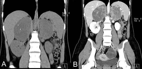
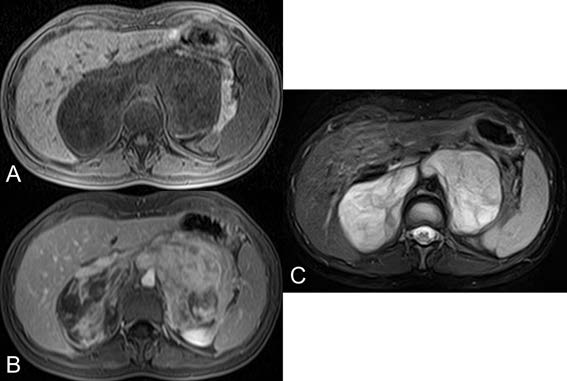
A 22-year-old female, previously healthy, without alcohol or smoking habits, was referred to our hospital after an emergency appendectomy in October 2019 at Centro Hospitalar da Cova da Beira (CHCB). During appendectomy, surgeons found a retroperitoneal mass. There was no previous knowledge of this retroperitoneal mass, and the patient had never had any abdominal symptoms before. Abdominal computed tomography was initially performed to better characterize the mass, followed by magnetic resonance. What is the most probable diagnosis?

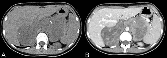
Figure 1: Axial soft tissue window, 1.2 mm slice thickness, non-enhanced CT (A), and contrast enhanced CT (B). 

Figure 2: coronal soft tissue window, non-enhanced CT (A), and contrast-enhanced CT (B). 

Figure 3: axial T1 weighted (A), axial T1 weighted contrast-enhanced (B), and T2 FS weighted (C) MRI 

Creative Commons License This is an open-access article distributed under the terms of the Creative Commons Attribution License