SciELO - Scientific Electronic Library Online

 
vol.34 número3Carcinoma dos Ductos Salivares da Glândula Parótida: um Cancro Raro e uma Potencial Armadilha SinistraPET com 2-[18F]-FDG no Diagnóstico Diferencial entre Doença de Alzheimer e Demência Frontotemporal índice de autoresíndice de assuntosPesquisa de artigos
Home Pagelista alfabética de periódicos  

Serviços Personalizados

Journal

Artigo

Indicadores

Links relacionados

  • Não possue artigos similaresSimilares em SciELO

Compartilhar


Acta Radiológica Portuguesa

versão impressa ISSN 2183-1351

Acta Radiol Port vol.34 no.3 Lisboa dez. 2022  Epub 30-Dez-2022

https://doi.org/10.25748/arp.27307 

Casos Clínicos

Giant Colonic Diverticulum a Rare Complication of a Common Disease

Divertículo Cólico Gigante uma Complicação Rara de uma Doença Comum

1Serviço de Cirurgia Geral, Hospital de Braga, Braga, Portugal

2Serviço de Cirurgia Geral, Hospital Póvoa de Varzim e Vila do Conde, Póvoa de Varzim, Portugal

3Serviço de Cirurgia Geral, Hospital Santa Maria Maior, Barcelos, Portugal


Abstract

An 80-year-old patient presented to the emergency room with a bowel obstruction and a tender epigastric mass. An abdominal computed tomography (CT) described a closed loop small bowel obstruction. An exploratory laparotomy revealed a giant sigmoid diverticulum, of 11cm in diameter, which was resected. The specimen was a true McNutt's type III diverticulum. The patient was discharged after a normal postoperative period and did not suffer any recurrence during a follow-up of more than 6 months. This is a rare complication of a common disease with non-specific presentation and not always accurately described by CT scan. Apart from intestinal volvulus, differential diagnoses include duplication cyst, a giant Meckel’s or duodenal diverticulum, pancreatic pseudocyst and pneumatosis cystoides intestinalis. Surgical resection is the recommended treatment to avoid perforation and peritonitis. Diverticulectomy is a safe option for a patient with acute presentation in the emergency room.

Keywords: Giant colonic diverticulum; Diverticular disease; Bowel obstruction; Computed tomography.

Resumo

Um doente de 80 anos recorreu ao serviço de urgência, em oclusão, com uma massa epigástrica dolorosa. A Tomografia computorizada (TC) indicava oclusão de delgado, em ansa fechada. Na laparotomia exploradora constatou-se um divertículo gigante do sigmóide, com 11cm de diâmetro, sem qualquer alteração a nível do intestino delgado. Procedeu-se a diverticulectomia. A histologia da peça revelou tratar-se de um divertículo verdadeiro tipo III, segundo a classificação de McNutt. O doente permaneceu assintomático, ao fim de mais de 6 meses de seguimento. Um divertículo gigante é uma complicação rara, de uma doença comum. A sua apresentação, inespecífica, nem sempre é esclarecida pelos achados da TC. Além de volvo intestinal, outros diagnósticos diferenciais incluem cistos de duplicação, divertículo gigante de Meckel ou duodenal, pseudocisto pancreático ou Pneumatose cística intestinal. O tratamento deve passar por resseção de forma a evitar perfuração e peritonite. A diverticulectomia é uma opção válida e menos invasiva.

Palavras-chave: Divertículo gigante; Doença diverticular; Oclusão intestinal; Tomografia computorizada.

Case Report/Case Presentation

This case reports an 80-year-old male patient with significant cardiac comorbidities including hypertension, type 2 diabetes and a previous ischaemic stroke. He reported epigastric pain, absence of passage of flatus and diminished elimination of faeces, within a period of 3 days. On physical examination, he was dehydrated, febrile, the abdomen was distended with hypoactive bowel sounds and a tender epigastric mass, without peritoneal signs.

The blood tests revealed leucocytosis and elevated C-reactive protein. An abdominal computed tomography showed an 11 cm air-filled central structure, apparently originating from the small bowel, described as a closed loop small bowel obstruction (Fig.1).

Figure 1: CT scan showed an 11 cm air-filled central structure 

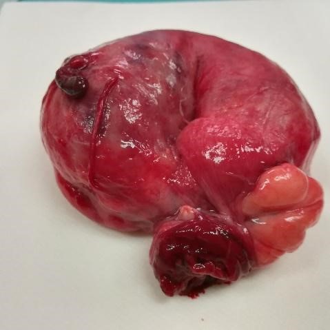
A laparotomy was performed and a large gas-filled diverticulum, at the antimesenteric border of the sigmoid colon, with an epiploon tourniquet at the diverticular neck was found (Fig. 2). It was associated with an extensive diverticulosis of the left colon. Diverticulectomy alone was safely performed (Fig.3).
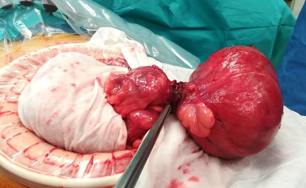
Figure 2: Giant colonic diverticulum, at the antimesenteric border of the sigmoid colon, with an epiploon tourniquet at the diverticular neck 

Figure 3: Surgical resection specimen 

The patient was discharged home after a normal postoperative period of 9 days. During the follow-up period of 6 months, he did not suffer any recurrence.

Discussion/Conclusion

A giant colonic diverticulum (GCD) is a rare complication of the common diverticular disease. Since it was first described in 19461,2, less than 200 cases have been reported in the literature worldwide.

A giant colonic diverticulum is 4 cm or greater in diameter and it can remain asymptomatic for a long period of time, until it reaches 7 cm or more3. In order to explain the size of the diverticula, it is postulated that the fibrous neck above the opening of the diverticulum acts as a one-way valve which allows air entry but not its exit1,2,3,4,5. It has also been attributed to the action of gas-forming organisms1,6. In spite of reports of GCD without additional diverticula, diverticulosis is present in 90% of cases and approximately 81% - 90% involve the sigmoid colon1,2,3,4,5.

Histologically, McNutt described three types of diverticula. Type I is a pseudodiverticulum with an out-pouching of mucosa and submucosa that protrudes through a defect in the colonic wall. Type II is not a diverticulum, but a walled-off abscess cavity due to perforation of a diverticulum that remains in contact with the lumen of the colon; the wall is made of fibrous scar tissue that enlarges to giant size. Type III is a true diverticulum, in which the wall contains all layers of the colonic wall1,2,3,4,5. Type III is the rarest described in reviews, found in only 12% of cases2.

Its presentation is generally non-specific. One third of patients present acutely with abdominal pain1 associated with fever, nausea, vomiting and rectal bleeding. As a chronic or sub-acute manifestation, symptoms include intermittent abdominal discomfort, bloating, and constipation, which may be associated with a palpable and soft abdominal mass, with variations in size. The presence of an intermittently palpable mass is responsible for the designation of ‘phantom tumour’, associated with GCD2,6. Approximately 10% of the patients are asymptomatic, but the mass can be detected either upon examination, or as an air-filled cystic image observed in an imaging procedure1,5. Diagnosis is based predominantly on abdominal computed tomography, which is the most accurate and recommended examination. The CT demonstrates a smooth-walled structure filled with stool and gas, that communicates with the colonic lumen1. Although the rarity of GCD can lead to the misdiagnosis of large bowel volvulus, as seen in the present case report6, other differential diagnoses include a duplication cyst, a giant Meckel’s diverticulum, duodenal diverticula, infected pancreatic pseudocyst and pneumatosis cystoides intestinalis7,8.

Due to their propensity to cause complications, surgical resection is the recommended treatment, even for asymptomatic patients3. The complications range from peritonitis, caused by the perforation of the GCD, abscess formation, intestinal obstruction, volvulus and infarction. The majority of patients are treated with colonic resection with en bloc resection of the diverticulum. However, Hartmann’s procedure and diverticulectomy are also performed. Non-surgical treatments might be indicated for high-risk patients, or those refusing surgery. Percutaneous drainage, stent placement, and antibiotics are typically followed by delayed elective segmental colectomy1,2.

The presented case describes an elderly patient, without previous manifestation of diverticular disease, in which the computed tomography did not reveal the accurate diagnosis. The acute presentation required urgent surgery and revealed a CGD of 11 cm in diameter and not a closed loop small bowel obstruction. Type III is the rarest described in reviews, with a proportion of 12% of cases found. Bearing in mind the age and comorbidities of the patient, diverticulectomy was a safe and adequate choice for this urgent procedure. The patient remained asymptomatic during the follow-up, with no evidence of recurrence.

References

1. Nigri G, et al. Giant colonic diverticulum, clinical presentation, diagnosis and treatment: Systematic review of 166 cases. World J Gastroenterol. 2015;21:360-8. [ Links ]

2. Praveen BV, et al. Giant colonic diverticulum: an unusual abdominal lump. J Surg Educ. 2007;64:97-100. [ Links ]

3. Carr, J. Alfred. Case report of a giant colonic sigmoid diverticulum causing sigmoidvolvulus. International Journal of Surgery Case Reports. 2017;1:197-9. [ Links ]

4. Cronin TG Jr, Tway MS, Boraca CT. Recurrent giant air cyst of the colon. IMJ Ill Med J. 1981;160:40-2. [ Links ]

5. Pozo AC del, et al. A gas-filled abdominal cyst in an elderly woman: A giant colonic diverticulum case report. International Journal of Surgery Case Reports. 2016;24:104-7. [ Links ]

6. Zeina AR, et al. Giant colonic diverticulum: radiographic and MDCT Characteristics. Insights Imaging. 2015;6:659-64. [ Links ]

7. Beddy D, DeBlacam C, Mehigan B. An Unusual Cause of an Acute Abdomen-a Giant Colonic Diverticulum. J Gastrointest Surg. 2010;14:2016-7. [ Links ]

8 Thomas S, Peel RL, Evans LE et-al. Best cases from the AFIP: Giant colonic diverticulum. Radiographics. 2006;26:1869-72 [ Links ]

Ethical disclosures

Financing Support: This work has not received any contribution, grant or scholarship.

Received: June 02, 2022; Accepted: September 15, 2022

Address Isabel Maria Lucas Marques, Serviço de Cirurgia Geral, Hospital de Braga, Sete Fontes - São Victor, 4710-243 Braga, Portugal, e-mail: isabel.m.marques@hb.min-saude.pt

Conflicts of interest: The authors have no conflicts of interest to declare.

Confidentiality of data: The authors declare that they have followed the protocols of their work center on the publication of data from patients.

Protection of human and animal subjects: The authors declare that the procedures followed were in accordance with the regulations of the relevant clinical research ethics committee and with those of the Code of Ethics of the World Medical Association (Declaration of Helsinki).

© Author(s) (or their employer(s)) and ARP 2022. Re-use permitted under CC BY-NC. No commercial re-use.

Creative Commons License This is an open-access article distributed under the terms of the Creative Commons Attribution License