SciELO - Scientific Electronic Library Online

 
vol.35 issue1Hepatic Artery Pseudoaneurysm After Liver Transplantation - Endovascular Treatment with Stent GraftBladder Rhabdomyosarcoma in an Infant with Schimmelpenning Syndrome author indexsubject indexarticles search
Home Pagealphabetic serial listing  

Services on Demand

Journal

Article

Indicators

Related links

  • Have no similar articlesSimilars in SciELO

Share


Acta Radiológica Portuguesa

Print version ISSN 2183-1351

Acta Radiol Port vol.35 no.1 Lisboa Apr. 2023  Epub Apr 30, 2023

https://doi.org/10.25748/arp.26361 

Imagens de Interesse

Stump Appendicitis: A Challenging Diagnosis

Apendicite do Coto Apendicular: um Desafio Diagnóstico

1Department of Radiology, Hospital de Braga, Braga, Portugal

2Department of Radiology, Centro Hospitalar Universitário do Porto, Porto, Portugal


Abstract

Appendicitis of the appendicular stump is a rare surgical complication characterized by inflammation of the residual appendix.

A past history of appendectomy and abdominal pain with nonspecific characteristics are frequent causes of delayed diagnosis, which may be associated with serious complications, namely, intra-abdominal abscesses, perforations, peritonitis and septic shock.

Computed tomography (CT) plays a central role in timely diagnosis and in reducing the morbidity and mortality of this condition.

Thus, we present the case of a 35-year-old man who underwent appendectomy 8 months before, who presented at the emergency department with a 12-hour abdominal pain and the CT was essential for the diagnosis and treatment.

Key words: Cecal diseases; Appendix; Appendicitis; Appendectomy.

Resumo

A apendicite do coto apendicular representa uma complicação cirúrgica rara, caracterizada pela inflamação do apêndice residual.

A história pregressa de apendicectomia e a dor abdominal de características inespecíficas constituem causas frequentes de atraso do diagnóstico, podendo associar-se a complicações graves, nomeadamente, abcessos intra-abdominais, perfurações, peritonites e choque séptico.

A tomografia computorizada (CT) desempenha um papel central no diagnóstico atempado e na redução da morbi-mortalidade desta condição.

Assim, apresentamos o caso de um homem de 35 anos submetido a apendicectomia 8 meses antes, que recorre ao serviço de urgência por dor abdominal com 12 horas de evolução e em que a CT foi essencial no diagnóstico e tratamento.

Palavras-chave: Doenças cecais; Apêndice; Apendicite; Apendicectomia.

Case report

A 35-year-old man presented to the emergency department with a 12-hour history of pain that started in the epigastric area and then migrated to the right lower quadrant. He did not report vomiting, change in bowel habits or dysuria.

Physical examination showed axillary temperature, 38.10C; tenderness at palpation over McBurney´s point without rebound. The patient had been submitted to appendectomy 8 months earlier.

Blood tests showed leukocytosis (13.1 x 10⁹/ L; N: 4-11) with 76% neutrophilia and a sightly C-reactive protein increase (5.4 mg/dL; N: <3). Urinalysis was normal.

Abdominal ultrasound showed a noncompressible hypoechogenic oval mass, with surrounding hyperechogenic fat and small amount of fluid in the right iliac fossa (figure 1).
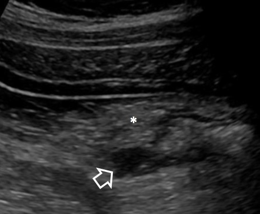
Figure 1: Abdominal ultrasound shows an oval hypoechoic formation arising from the base of the cecum (open arrow) and thickening of the surrounding mesenteric fat (*). 

CT scan with intravenous contrast of the abdomen and pelvis was performed, demonstrating a tubular structure with enhancing wall extending from the base of the cecum and stranding of the adjacent fat (figure 2), findings suggestive of stump appendicitis.
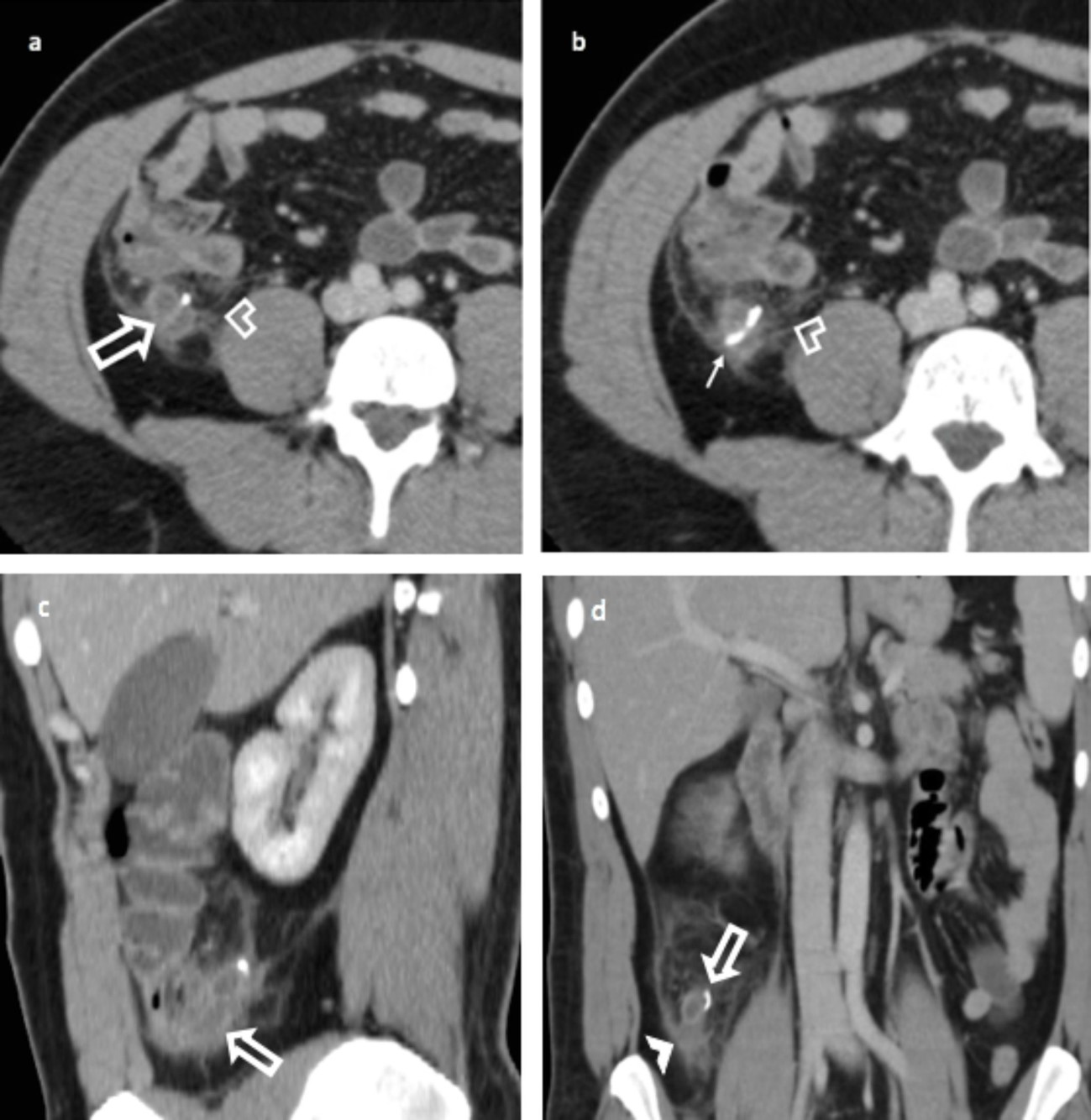
Figure 2: Axial contrast-enhanced CT scan series of abdomen (a, b) show enlarged appendiceal stump with enhancing wall (open arrow); thickening of the peritoneal reflection can also be seen (open arrowhead). Note staple line (b) from initial appendectomy (solid arrow). Sagittal (c) and coronal (d) reformatted images better depict the enlarged appendiceal stump arising from cecum (open arrowhead). There is stranding of adjacent mesenteric fat and local free fluid (solid arrowhead). Imaging also excludes free intraperitoneal air and abscess formation. 

The patient underwent exploratory laparotomy and acute inflammation and obstruction of the stump appendix by an appendicolith was confirmed in pathology. There was no perforation or abscess formation.

The patient did well and was discharged home two days after surgery.

Discussion

Appendectomy is the most common surgery worldwide. Stump apendicitis is a rare surgical complication (1/50 000 cases) characterized by inflammation of the appendiceal remnant after incomplete appendectomy. It is reported to have increased since the introduction of laparoscopic appendectomy.1

The remnant appendiceal greater than 5 mm in length is a risk factor for the formation of fecaliths and consequently stump appendicitis.2 The experience of the surgeon, the subserous or retrocecal position of appendix and an inadequate dissection technique are other known risk factors for stump appendicitis.2,3

The time to develop this complication is long, ranging from 9 weeks to 50 years and the signs and symptoms are similar to the classical acute appendicitis, leading to a delay in diagnosis due to the previous history of appendectomy.2

Unrecognition of this entity causes a delay in the diagnosis, leading to severe complications, such as abscess formation, high rates of perforation (70%) and septic shock.4,5

Abdominal ultrasonography and CT are useful tools for attempted diagnosis. Ultrasonography can demonstrate the appendiceal remnant, as a tubular structure extending from the right iliac fossa or retrocecal region to the cecum, the hyperechogenic of periappendiceal fat and free fluid, but it is operator dependent and it is limited by the patient biotype as well.

Abdominal CT scan is more specific than US in depicting the typical findings, being the exam modality of choice to characterize the complications, such abscesses, perforation and peritonitis.

Prompt suspicion and confirmation of the diagnosis with CT is therefore essential to avoid serious complications, improving morbidity and mortality.4

References

1. Aschkenasy MT, Rybicki FJ. Acute appendicitis of the appendiceal stump. J Emerg Med. 2005;28:41-3. [ Links ]

2. Durgun AV, Baca B, Ersoy Y, Kapan M. Stump appendicitis and generalized peritonitis due to incomplete appendectomy. Tech Coloproctol. 2003;7:102-4. [ Links ]

3. Dikicier E, Altintoprak F, Ozdemir K et al. Stump appendicitis: a retrospective review of 3130 consecutive appendectomy cases. World J Emerg Surg, 2018;13:22 [ Links ]

4. Awe JA, Soliman AM, Gourdie RW. Stump appendicitis: an uncompleted surgery, a rare but important entity with potential problems. Case Rep Surg. 2013;2013:972596. [ Links ]

5. Uludag M, Isgor A, Basak M. Stump appendicitis is a rare delayed complication of appendectomy: a case report. World J Gastroenterol. 2006;12:5401-3. [ Links ]

Ethical disclosures

Financing Support: This work has not received any contribution, grant or scholarship.

Received: February 01, 2022; Accepted: March 01, 2022

Address Ana Catarina Costa, Serviço de Radiologia, Hospital de Braga, Sete Fontes São Victor, 4710-243 Braga, Portugal, e-mail: anacatarinagcosta@gmail.com

Conflicts of interest: The authors have no conflicts of interest to declare.

Confidentiality of data: The authors declare that they have followed the protocols of their work center on the publication of data from patients.

Protection of human and animal subjects: The authors declare that the procedures followed were in accordance with the regulations of the relevant clinical research ethics committee and with those of the Code of Ethics of the World Medical Association (Declaration of Helsinki).

© Author(s) (or their employer(s)) and ARP 2023. Re-use permitted under CC BY-NC. No commercial re-use.

Creative Commons License This is an open-access article distributed under the terms of the Creative Commons Attribution License