SciELO - Scientific Electronic Library Online

 
vol.7 número1Paniculite Pancreática: Forma de Apresentação RaraTentativa de Suicídio Atípica com Laceração da Veia Jugular na Adolescência: A Propósito de um Caso Clínico índice de autoresíndice de materiabúsqueda de artículos
Home Pagelista alfabética de revistas  

Servicios Personalizados

Revista

Articulo

Indicadores

Links relacionados

  • No hay articulos similaresSimilares en SciELO

Compartir


Gazeta Médica

versión impresa ISSN 2183-8135versión On-line ISSN 2184-0628

Gaz Med vol.7 no.1 Queluz mar. 2020  Epub 07-Jun-2021

https://doi.org/10.29315/gm.v7i1.291 

Imagens Médicas

Recurrent Intussusception in Adolescence: A Rare Entity

Invaginação Recorrente na Adolescência: Uma Entidade Rara

Cátia Vilas Boas Leitão1 
http://orcid.org/0000-0003-1470-3148

Joana Tenente1 

Andreia Ribeiro2 

Isabel Pinto Pais2 

1 Resident of Pediatrics Department, Centro Hospitalar de Vila Nova de Gaia/Espinho, Vila Nova de Gaia, Portugal.

2 Pediatric Gastroenterology Unit, Pediatrics Department, Centro Hospitalar Vila Nova de Gaia/Espinho, Portugal.


Keywords: Adolescent; Intussusception

Palavras-chave: Adolescente; Invaginação Intestinal

A healthy 16-year-old male presented to the emergency department with vomiting and pain in the right iliac fossa for the previous 12 hours, with no other symptoms. On the physical examination the patient had pain in the right iliac fossa and localized rebound tenderness. Na abdominal ultrasound showed a small bowel intussusception with approximately 4.9 cm. Blood tests were normal, stool cultures and virology were negative. The intussusception reduced spontaneously in 24 hours. One week later the patient had the same complaints and a new abdominal ultrasound confirmed the recurrence of the intussusception (Fig. 1). The abdominal and pelvic computed tomography scan showed no other alterations.

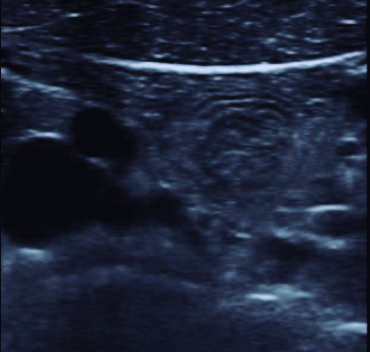
Figure 1: Ultrassound transverse image of a target shaped lesion in the right iliac fossa, classical described as the “doughnut” or “target” sign, corresponding to the concentric rings of bowel Wall (inside the red square). 

The intussusception reduced spontaneously. The investigation was negative for intestinal parasitosis, celiac disease and ectopic gastric mucosa. A video capsule endoscopy revealed small nodules in the distal ileum related to lymphoid hyperplasia (Fig. 2). After 2 years of follow-up, there were no signs of recurrence.

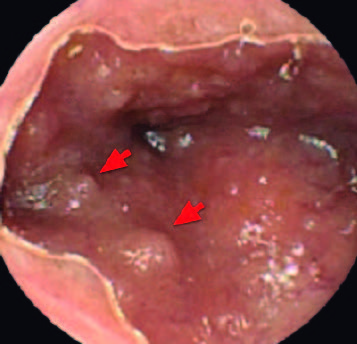
Figure 2: Video capsule endoscopy image showing small mucosa nodules (red arrows) in the terminal ileum, suggestive of nodular lymphoid hyperplasia 

.

Intussusception is a common cause of intestinal obstruction in infants and children.1-3 In older children, especially adolescents, it is less frequent and has an atypical presentation in most cases.1,2 Although the majority of pediatric cases are idiopathic, in 25% a pathological lead point (PLP) can be determined, such as Meckel’s diverticulum, intestinal duplication, lymphoid hyperplasia, intestinal polyps, vascular malformations or malignancies.1,4 Clinical suspicion is extremely important at every age, since the delayed diagnosis may lead to intestinal ischemia, perforation and peritonitis.1,3

Recurrent intussusception occurs in approximately 10% of the cases. The risk factors have not been clearly defined, but older age (>1-2 years), absence of vomiting and PLP can be predictors of recurrent intussusception.3,5

Intestinal lymphoid hyperplasia is a benign condition and may act as a PLP for intussusception.4 A course of oral steroids may be used in cases of multiple recurrences; however, this approach needs more studies to be recommended.6

References

1. Marsicovetere P, Ivatury SJ, White B, Holubar S. Intestinal intussusception: etiology, diagnosis, and treatment. Clin Colon Rectal Surg. 2017; 30: 30-9. doi: 10.1055/s-0036-1593429. [ Links ]

2. Mandeville K, Chien M, Willyerd F, Mandell G, Hostetler M, Bulloc B. Intussusception: clinical presentations and imaging characteristics. Pediatr Emerg Care. 2012; 28: 842-4. [ Links ]

3. Guo W, Hu Z,Tan Y, Wang J. Risk factors for recurrent intussusception in children: a retrospective cohort study. BMJ Open 2017;7:e018604. doi:10.1136/bmjopen-2017-018604. [ Links ]

4. Ntoulia A, Tharakan SJ, Reid JR, Mahboubi S. Failed intussusception reduction in children: correlation between radiologic, surgical, and pathologic findings. AJR Am J Roentgenol. 2016;207:424. [ Links ]

5. Ye X, Tang R, Chen S, Lin Z and Zhu J. Risk Factors for Recurrent Intussusception in Children: A Systematic Review and Meta-Analysis. Front. Pediatr. 2019; 7:145. doi: 10.3389/fped.2019.00145. [ Links ]

6. Gluckman S, Karpelowsky J, Webster AC, McGee RG. Management for intussusception in children. Cochrane Database Syst Rev. 2017;6:CD006476. doi: 10.1002/14651858.CD006476.pub3. [ Links ]

Fontes de financiamento: Não houve qualquer fonte de financiamento na realização do presente trabalho.

Confidencialidade dos dados: Os autores declaram ter seguido os protocolos da sua instituição acerca da publicação dos dados de doentes.

Consentimento: Consentimento do doente para publicação obtido.

Proveniência e revisão por pares: Não comissionado; revisão externa por pares

Financial support: This work has not received any contribution, grant or scholarship

Confidentiality of data: The authors declare that they have followed the protocols of their work center on the publication of data from patients

Patient consent: Consent for publication was obtained

Provenance and peer review: Not commissioned; externally peer reviewed

Received: December 14, 2019; Accepted: March 26, 2020; Published: March 31, 2020

Corresponding Author/Autor Correspondente: Cátia Vilas Boas Leitão [catiavbl@gmail.com] Rua Francisco Sá Carneiro, s/n, 4400-129 Vila Nova de Gaia, Portugal ORCID iD: 0000-0003-1470-3148

Conflitos de interesse:

Os autores declaram não ter qualquer conflito de interesse na realização do presente trabalho.

Conflicts of interest:

The authors declare that they have no conflicts of interest.

Creative Commons License This is an open-access article distributed under the terms of the Creative Commons Attribution License