SciELO - Scientific Electronic Library Online

 
vol.8 número2Migração Intra-Abdominal de Sistema Intrauterino Após Perfuração Uterina índice de autoresíndice de assuntosPesquisa de artigos
Home Pagelista alfabética de periódicos  

Serviços Personalizados

Journal

Artigo

Indicadores

Links relacionados

  • Não possue artigos similaresSimilares em SciELO

Compartilhar


Gazeta Médica

versão impressa ISSN 2183-8135versão On-line ISSN 2184-0628

Gaz Med vol.8 no.2 Queluz jun. 2021  Epub 01-Set-2021

https://doi.org/10.29315/gm.v8i2.434 

Imagens Médicas

Perforated Diverticulitis Complicated by Pylephlebitis and Splenic Infarction

Diverticulite Perfurada Complicada com Pileflebite e Enfarte Esplénico

Catarina Janicas1  , Interno de Formação Específica de Radiologia
http://orcid.org/0000-0002-3989-0931

David Campos-Correia1  , Interno de Formação Específica de Radiologia

Ana Paula Vasconcelos2  , Assistente Hospitalar Graduado de Radiologia

1. Interno de Formação Específica de Radiologia, Serviço de Radiologia, Centro Hospitalar de Lisboa Ocidental, Lisboa, Portugal.

2. Assistente Hospitalar Graduado de Radiologia, Serviço de Radiologia, Centro Hospitalar de Lisboa Ocidental, Lisboa, Portugal.


Keywords: Diverticulitis; Splenic Infarction; Thrombophlebitis; Tomography, X-Ray Computed

Palavras-chave: Diverticulite; Enfarte do Baço; Tomografia Computorizada; Tromboflebite

A 64-year-old male presented to our emergency department with a 6-day history of generalized malaise, worsened by left lower back pain and anorexia for the last 2 days. Other symptoms were denied, and analytical evaluation only showed leucocytosis and elevated C-reactive protein. Contrast-enhanced computed tomography (CE-CT) was performed (Fig. 1), in which a thickened segment of sigmoid colon with diverticula, peri-colonic fat stranding and a small amount of extra-luminal gas were seen, in keeping with perforated diverticulitis. It also revealed air thrombi along the sigmoid veins and the inferior mesenteric vein, as well as a wedge-shaped hypoenhancing area in the spleen, interpreted as splenic infarction secondary to pylephlebitis.

Figure 1: Perforated diverticulitis complicated by pylephlebitis and splenic infarction. Admission contrast-enhanced CT reveals focal fat stranding and a small amount of extra-luminal gas (arrow, a) adjacent to a thickened segment of sigmoid colon with diverticula, suggesting perforated diverticulitis. Several filling defects, compatible with air thrombi, are seen along the densified sigmoid veins (arrowheads, a) and inferior mesenteric vein (arrowheads, b). The spleen has a wedge-shaped hypoenhancing area (asterisk, c), in relation to infarction caused by the air thrombi already described. There are neither local/distant abscesses nor signs of generalized peritonitis. 

Despite initially showing complete clinical response under broad-spectrum antibiotics, the patient relapsed with fever and left lower back pain. CE-CT was again performed (Fig. 2), showing evolution of the previously documented splenic infarct into spontaneous splenic rupture. He was submitted to emergent splenectomy and peritoneal lavage, with posterior clinical recovery and discharge.
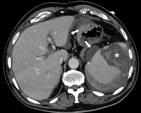
Figure 2: Spontaneous splenic rupture. In the contrast-enhanced CT performed twelve days later, the spleen is enlarged and an extensive intraparenchymal and subcapsular hematoma has developed in the infarcted area (asterisk). A small hemoperitoneum with no detectable foci of contrast extravasation (arrows) is also seen. 

Acute diverticulitis is a well-documented cause of acute abdominal pain, often complicated by intra-abdominal abscess, fistulisation and perforation.1 Very rarely, due to high endoluminal pressure and bacterial transmigration, septic or gas emboli are released from the affected area and cause ascending venous thrombosis (pylephle-bitis).2 Mesenteric pylephlebitis may cause segmental bowel ischemia or, through embolization along the splenic and portal veins, distant abscess formation and infarction.1,2 Exceedingly rarely, pylephlebitis of the inferior vena cava occurs, resulting in septic pulmonary embolism.1,3

Because clinical presentation is often unspecific,2 CE-CT is the modality of choice. It allows not only visualization of thrombi and estimation of disease extent, but also identification of its underlying cause.1,2 Although the prognosis of gas in the venous system and spontaneous splenic rupture is generally very poor, it tends to be more favourable in cases of benign etiology as diverticulitis.1,2,4

Referências

1. Onur MR, Akpinar E, Karaosmanoglu AD, Isayev C, Karcaaltincaba M. Diverticulitis: a comprehensive review with usual and unusual complications. Insights Imaging. 2017;8:19-27. doi: 10.1007/s13244-016-0532-3. [ Links ]

2. Rose J, Khoubyari R, McClenathan J. Diverticulitis as a cause of septic thrombophlebitis: a literature review. IJCRI. 2011;2:28-33. doi: 10.5348/ijcri20111277CR7. [ Links ]

3. Isik M, Cinar E, Kizilarslanoglu MC, Ozbek E, Etgul S, Kiraz S. A confusing case: pulmonary lesions including cavities, isolated left heart endocarditis and inferior vena cava thrombosis in a patient with perforated diverticulitis. Rheumatol Int. 2013;33:2179-81. doi: 10.1007/s00296-012-2424-2. [ Links ]

4. Tonolini M, Ierardi AM, Carrafiello G. Atraumatic splenic rupture, an underrated cause of acute abdomen. Insights Imaging. 2016;7:641-6. doi: 10.1007/s13244-016-0500-y. [ Links ]

Responsabilidades éticas

Fontes de financiamento: Não existiram fontes externas de financiamento para a realização deste artigo

Confidencialidade dos dados: Os autores declaram ter seguido os protocolos da sua instituição acerca da publicação dos dados de doentes

Consentimento: Consentimento do doente para publicação obtido

Proveniência e revisão por pares: Não comissionado; revisão externa por pares

Ethical disclosures

Financing support: This work has not received any contribution, grant or scholarship

Confidentiality of data: The authors declare that they have followed the protocols of their work center on the publication of data from patients

Patient consent: Consent for publication was obtained

Provenance and peer review: Not commissioned; externally peer reviewed

Received: March 05, 2021; Accepted: March 30, 2021; Published: June 30, 2021

Catarina do Carmo Janicas [ccjanicas@hotmail.com] Estrada Forte do Alto Duque, 1449-005 Lisboa, Portugal ORCID iD: 0000-0002-3989-0931

Conflitos de interesse:

Os autores declaram a inexistência de conflitos de interesse na realização do presente trabalho.

Conflicts of interest:

The authors have no conflicts of interest to declare.

Creative Commons License This is an open-access article distributed under the terms of the Creative Commons Attribution License