


 Inglês (pdf)
 Artigo em XML
 Referências do artigo
 Enviar este artigo por email
 Citado por SciELO
  Similares em
SciELO
Similares em
SciELO 
 Permalink
Introduction
Parsonage-Turner syndrome (PTS) is defined as severe acute-onset shoulder pain followed by severe muscle weakness in the distribution of one or more nerves of brachial plexus.1,2 The pain often awakes the patient in the middle of the night, typically lasts two to three weeks and then resolves being followed by severe muscle weakness.2,3
PTS is also referred to as acute brachial neuritis or neuralgic amyotrophy.4 It was initially described in 1897.5) In 1948 Parsonage-Turner reported the first large series of patients.1
Classically, Parsonage-Turner syndrome is described as a rare disease, its incidence being 1-3 per 100 000.2,6 However, it is now understood that the lack of PTS recognition reduces its true incidence.7 In fact, a recent prospective study reported an incidence of 1:1000.8 Male:female ratio is 2:1.9 The average age of onset is 40 years-old in the idiopathic form and 25 years-old in the inherited form.3
PTS etiology is unknown, and it likely represents a complex syndrome comprising different underlying mechanisms, phenotypes, and prognoses.2,10) It is widely believed that PTS initiates from an autoimmune response after an illness or environmental factor such as strenuous activity, vaccination, surgery, or childbirth.9,11 The most common trigger is a preceding viral infection occurring one to two weeks before shoulder symptoms develop, and is found in up to 55% of PTS.9 In approximately half of the cases the patient is unable to report any of the known risks.10 Other authors believe that there might be a vasculitis/ischemia component which better explains the acute onset of pain.2,11 Finally, some refer a mechanical component - traction and compression of the nerves.11) Most PTS are idiopathic.9 The inherited form, also called hereditary neuralgic amyotrophy, is rare and has been associated with alterations in the SEPT9 gene.9 Clinically, it is similar to the idiopathic PTS, but attacks tend to occur more frequently.9
Diagnosis is challenging since pain presentation may not always be typical, major risk factors may not always be present, and weakness is often delayed or initially masked by the severe pain.7 A careful shoulder examination looking for single and multiple mononeuropathies affecting the proximal musculature is crucial.3 The affected muscles do not share root innervation, which would imply radiculopathy instead.3 Scapular dyskinesia is frequent and therefore should be tested.3 Classically, diagnosis is mainly based on clinical observations.2 Electromyography is useful in identifying the location of a suspected neurological deficit, in confirming the suspicion of PTS (as opposed to a more global brachial plexopathy or a cervical radiculopathy), and in monitoring the course of the disease.12,13 Typical electromyography findings include widespread/complete denervation in the involved muscles.14 Motor and sensory nerve conduction velocities and distal latencies on the distal upper limb are usually normal, as PTS classically affects the proximal muscles of the upper limb.14 Magnetic resonance imaging (MRI) helps excluding other causes of shoulder pain such as rotator cuff tear, impingement syndromes, labral tears, spinoglenoid and suprascapular notch masses.14 It also detects signal abnormalities in the shoulder girdle musculature related to denervation such as diffuse increased T2-weighted signal, which may occur without a T1-weighted signal change or, in the subacute and chronic phases of denervation, T2-weighted signal abnormalities and muscular atrophy.14 Recently, improved imaging methods in MRI and high-resolution ultrasound have led to the identification of structural peripheral nerve lesions, most notably hourglass-like constrictions.10,15 These findings are considered pathognomonic and have led to more accurate diagnosis through high-resolution imaging.9,15,16
Traditionally, PTS prognosis is considered good, with full recovery occurring in 80%-90% of patients, two to three years following the onset of symptoms.11 Conversely, recent reports have shown that many patients end up with residual pain or decreased exercise tolerance in the affected limb.17 Two years after onset, approximately 1/4 to 1/3 of the patients still suffer from pain and muscle fatigue, and half of the patients either need to change their profession or are unable to work at all.8,17 Recovery is usually slow, lasting months to years and can be monitored through clinical examination and serial electromyograms.9,12
Previously, treatment options were confined to conservative measures, namely early administration of corticosteroids, appropriate pain management and physical therapy to cope with muscle weakness.9,18 More recently, peripheral nerve surgery has been shown to be a valuable treatment option in the cases of PTS with identified structural peripheral nerve lesions and no signs of clinical recovery after three months.9
The authors felt it would be important to revisit this subject since Parsonage-Turner syndrome is severely underdiagnosed in clinical practice, despite an actual estimated incidence of 1/1000 per year, and considering that prompt diagnosis is key to optimal management and patient’s reassurance.8
Case Reports
Four patients, two woman and two men, were diagnosed with Parsonage-Turner Syndrome. Ages ranged from 46 to 84 years old. In the four cases, diagnosis was established at least one month after the onset of symptoms. All patients visited more than one doctor until the diagnosis was reached. All patients were right-hand dominant, and the right shoulder was the one involved. Pain subsided after three to eight days. All patients recovered, at least partially, with conservative treatment, namely pain management and rehabilitation. Recovery took between six months and 15 months and was monitored through clinical evaluation and electromyograms every six months. At last follow-up, all patients were satisfied.
Case 1
A 46-year-old female flight attendant presented to our shoulder unit with a 1.5-month history of right shoulder pain and progressive weakness. The pain, located on the supraspinatus fossa, was severe (intensity of 8/10 on VAS), had an acute onset with no previous trauma or intense activity. However, she claimed that she had just recovered from a viral upper respiratory tract infection before shoulder symptoms developed. The pain significantly improved over time, but muscle weakness progressed. On initial clinical examination deltoid and supraspinatus fossa atrophy was evident. Right shoulder passive range of motion was unchanged, but right unilateral weakness on abduction and external rotation were noted.
Radiographs, ultrasound, and blood analysis were normal. MRI of the right shoulder showed supraspinatus and infraspinatus atrophy, with no signs of tear. Electromyogram confirmed a severe neuropathy of the suprascapular nerve and a moderate neuropathy of the axillary nerve. The patient underwent physical therapy, and six months later improvement of abduction and external rotation strength was evident, but still inferior to the healthy shoulder (4/5). At last follow-up, three years post-injury, she demonstrated full, pain-free shoulder range of motion, with 5/5 rotator cuff strength (Fig. 1). She returned to work and was able to resume sports practice (yoga) without limitations.
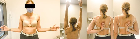
Figure 1: At last follow-up, three years post-injury, patient 1 demonstrated full, pain-free shoulder range of motion.
Case 2
A 50-year-old woman presented to our shoulder unit complaining of a 2-week right shoulder pain and weakness. Trauma, previous shoulder problems and recent infections were denied. Rhomboid muscles atrophy and medial winging of the scapula were evident upon inspection. The patient demonstrated a significant decrease in her right shoulder range of motion: 170º of forward elevation and abduction, 60º of external rotation, and internal rotation to the level of the 7th dorsal vertebra. Supraspinatus, infraspinatus, subscapularis, and trapezius strength was preserved.
Electromyogram showed a severe lesion of the long thoracic nerve and moderate lesion of the musculocutaneous nerve, supporting the Parsonage-Turner syndrome diagnosis. She was then referred to physical therapy with neurostimulation. Five months later, a control electromyogram revealed signs of improvement of the neuromotor pattern. At last follow-up (1.5-year post-injury), she presented full range of shoulder motion, and reported no pain apart from occasional minor discomfort (intensity of 1/10 on VAS) after specific movements during sports. The patient had already and uneventfully resumed work and was very satisfied.
Case 3
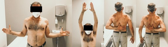
An otherwise healthy 59-year-old policeman presented to our office complaining of right shoulder pain and weakness. One month beforehand, he had woken up in the middle of the night with a sudden “terrible pain” in the right shoulder. In the emergency room, he denied a known injury or precipitating event and had no history of previous shoulder problems. He was given opioids with little pain relieve. He was then discharged and referred to our shoulder unit. On initial physical examination in clinic, one month after symptoms onset, he presented weakness of the right shoulder muscles, excluding trapezius and subscapularis. Muscle function distal to elbow was also preserved. Both passive motion of the right shoulder and radiographic exams were normal. An MRI of the right shoulder showed no evidence of rotator cuff tearing nor other distinct abnormalities. The clinical diagnosis of Parsonage-Turner syndrome was confirmed by electromyography which revealed brachial plexitis involving C5, C6 and C7 roots. The patient completed an 8-month period of physical therapy. He was unable to work for approximately one year. Three years post-injury, the patient presented full, pain-free shoulder range of motion and strength (5/5) and reported successfully resuming work and sports (Fig. 2). The patient was very satisfied.
Case 4
An 84-year-old man presented with a 5-month history of right pseudoparalytic shoulder. Six months before, he had suffered a stroke and the limitation of motion of the right shoulder progressed since then. The diagnosis was established approximately 5-6 months after the onset of symptoms. On examination, severe muscle atrophy and weakness of the supraspinatus, infraspinatus and deltoid were obvious. An MRI of the right shoulder revealed a partial tear of the infraspinatus with no muscle atrophy or tendon retraction, and grade I supraspinatus atrophy. At this point, differential diagnosis included brachial plexus, suprascapular and/or axillary lesions. Electromyogram confirmed severe partial lesion of the brachial plexus involving the superior trunk. Physical therapy was prescribed. Five months later, control electromyogram showed some reinnervation of the superior trunk of the brachial plexus, however, pseudoparalysis of the shoulder showed little improvement.
One year later, a new electromyogram showed even more reinnervation of the superior trunk and an improved range of motion was observed: 120º of forward flexion and abduction, 45º of external rotation and internal rotation to the level of the sacroiliac joint. He completed 15 months of physical therapy. At last follow-up (seven years post-injury), despite not having fully recovered, the patient was satisfied with the results, since pain-free shoulder motion needed for his daily life activities was regained.
Discussion
Involvement of the suprascapular, axillary, long thoracic, and musculocutaneous nerves and the superior trunk of the brachial plexus was detected through electromyography in the presented patients. Most commonly involved nerves are the suprascapular nerve that innervates the supraspinatous and infraspinatous musculotendinous units, responsible for forward elevation/abduction and external rotation, respectively; the long thoracic nerve which innervates the serratus anterior muscle, responsible for protraction and stabilization of the scapula (winging of the scapula); the anterior interosseous nerve, motor nerve that rises from the median nerve, and is responsible by flexion of the distal interphalanx of the thumb and second finger.4) Less commonly affected nerves include axillary nerve (deltoid, teres minor), radial/posterior interosseous nerve (extension of the elbow and wrist and fingers), musculocutaneous nerve (coracobrachialis, but biceps and brachialis sparing) and phrenic nerve (7% of PTS).4
The four patients presented with the typical sudden-onset pain in the shoulder region, followed by weakness of muscles in the shoulder and/or arm. Numerous reports have been published on the clinical spectrum of PTS.11 Initially, an hyperalgesic phase is characterized by severe pain and might persist from hours to 1-4 weeks.2 In the presented cases, pain resolved in three to eight days. Motor deficit with muscle weakness and atrophy are the main symptoms of the neuropathy, in contrast with the rare sensitive symptoms.2 A trigger event could not be found in most patients, except for case 1 who reported a preceding viral infection, which is the most common trigger.
Before diagnosis was established, all patients had had previous medical assistance elsewhere reporting the same symptoms. The idea that PTS is a rare disorder has recently been refuted as an annual incidence of 1/1000 has been reported in primary care, following a specific training of general practitioners on how to diagnose PTS.7 These recent reports suggest that PTS is severely underdiagnosed in day-to-day clinical practice.9) Therefore, it is of utmost importance to be aware of this pathology. Alfen et al8 confirmed PTS was poorly known, which justifies why it is diagnosed, in three of four cases, 28 weeks after disease onset (mean 44 weeks).
Recovery is assessed through muscle function which can be determined by serial physical examination and electromyography.7 Recently, hourglass-like fascicular constrictions and torsions of brachial plexus nerves on MRI have been found in PTS.10,19 Imaging is important to rule out other possible causes as extrinsic mass compression, assess muscle denervation edema pattern (bright signal) and evaluate for hourglass-like nerve constrictions (HGCs).20 HGCs in the form of “Bullseye sign” of the nerve, identified on cross-sectional MRI image immediately proximal to the site of constriction, have been seen in patients with PTS and confirmed surgically.20 These are potentially unique biomarkers of PTS.20
Opioids and non-steroidal anti-inflammatory drugs are often not helpful in reducing severe initial pain.7 Steroids have conventionally been prescribed as it has been reported they might shorten duration of pain and improve functional recovery.7 Physical therapy is considered the most effective treatment after signs of neurologic recovery.7 In the early phases of the disease, it might not be of dramatic benefit because frequently there are no electric impulses.2,7 Intravenous immunoglobulin (IVIG) has been proposed to have a possible benefit in patients with recurrent post-procedural attacks.17 There are no randomized controlled trials on how these medications may affect the disease course.17 For the patients with none or minimal recovery, surgical neurolysis of the constriction can be performed.18,21
Traditionally, PTS is thought to have a very favorable outcome, with nearly full recovery in 90% of the patients, after 1-3 years.9 Tsairis et al22 reported excellent functional recovery (36% at 1 year, 75% at 2 years and 89% at 3 years). Feinberg et al12 reported 80% of early axonal regeneration, 50% having full neurological recovery by 1 year. Suprascapular, spinal accessory, and axillary nerves recovered fastest.12 Long thoracic and anterior interosseous nerves were slower.12 Three of the four presented patients had virtually complete recoveries and are very satisfied. Patient 4 never fully recovered; however, his stroke sequelae are a confounding factor. Further treatment was not provided as the patient is a low-demanding 84-year-old man who recovered suitable function for his daily life activities. Recently, reports have shown fewer promising results. Van Alfen et al3) followed 246 patients for a minimal period of three years and observed only 8% had full recovery, 2/3 had persistent pain and paresis and >25% could not return to work. It is now assumed that most patients never achieve full recovery.9
This paper reports an illustrative case series of a condition that is frequently underdiagnosed and needs to be reminded in the differential diagnosis of the painful shoulder, especially if muscle weakness is present. Careful clinical examination and diagnosis support through electromyography improve opportune management and patient reassurance.
Conclusion
Parsonage-Turner syndrome is not as rare as previously assumed and is often underdiagnosed. It is therefore critical to be familiar with this condition. Overall, PTS is a self-limited idiopathic condition, and its core treatment is supportive. Most cases will not need further intervention than pain management and rehabilitation. A correct diagnosis based on the clinical findings and supported by electromyography and MRI is key and patient’s reassurance is required, considering recovery is usually slow.

