SciELO - Scientific Electronic Library Online

 
vol.9 número2Macroglossia Isolada ou Algo Mais?A Importância do Diagnóstico e Tratamento da Depressão Major em Doentes Oncológicos em Cuidados Paliativos índice de autoresíndice de materiabúsqueda de artículos
Home Pagelista alfabética de revistas  

Servicios Personalizados

Revista

Articulo

Indicadores

Links relacionados

  • No hay articulos similaresSimilares en SciELO

Compartir


Gazeta Médica

versión impresa ISSN 2183-8135versión On-line ISSN 2184-0628

Gaz Med vol.9 no.2 Queluz jun. 2022  Epub 30-Jun-2022

https://doi.org/10.29315/gm.v9i2.590 

Imagens Médicas

Pneumomediastinum and Pneumothorax in a Neonate with Meconium Aspiration Syndrome

Pneumomediastino e Pneumotórax em Recém-Nascido com Síndrome de Aspiração Meconial

1. Serviço de Pediatria, Hospital Pediátrico de Coimbra, CHUC, Coimbra, Portugal.

2. Serviço de Neonatologia, CHUC, Coimbra, Portugal.


Keywords: Infant, Newborn; Meconium Aspiration Syndrome/complications; Mediastinal Emphysema; Pneumothorax

Palavras-chave: Enfisema Mediastínico; Pneumotórax; Recém-Nascido; Síndrome de Aspiração Meconial/complicações

A term neonate born through meconium-stained amni- otic fluid from instrumented delivery, (Apgar score 7/8), without need for resuscitation, presents with grunting, cyanosis and respiratory distress by the 5th minute of life, with pulse oximetry revealing a 70%-75% saturation which resolved with oxygenotherapy.

The neonate was admitted to the neonatal intensive care unit (NICU), requiring oxygenotherapy (maxi- mum FiO2 40%, incubator environmental servo-oxy- gen delivery) for a saturation >95%. Initial bloodwork included a venous blood gases analysis (pH 7.26, pCO2 49.9 mmHg, HCO3- 22.4 mmol/L, BE -4.7 mmol/L, lac- tate 4.3 mmol/L), non-elevated C-reactive protein and a normal hemogram.

A chest x-ray showed a small pneumothorax (left lung apex) and left hemithorax radiolucency (outlining the mediastinal structures) with extrapleural air sign, suggesting a large pneumomediastinum. On the right hemi- thorax, diffuse, asymmetric, patchy pulmonary opacities mixed with focal overinflation areas are suggestive of meconium aspiration syndrome (MAS) (Fig. 1).

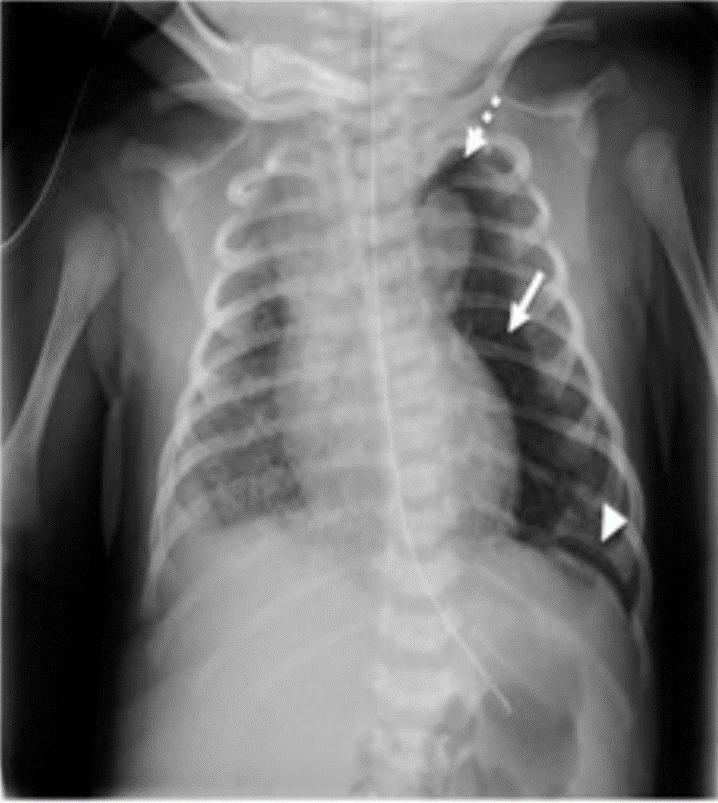
Figure 1: Chest x-ray at admission in NICU with a large pneumomediastinum (arrow) in the left hemithorax, with an extrapleural air sign (arrowhead) and a small linear pneumothorax (dotted arrow) on the left apex. Also visible are diffuse and asymmetric patchy opacities, mixed with focal areas of overinflation on the right pulmonary parenchyma, suggestive of MAS. 

Oxygen was successfully weaned off, with no signs of respiratory distress by the second day. A subsequent chest x-ray on the sixth day, showed complete reabsorption of the pneumomediastinum and pneumothorax (Fig. 2), after which the neonate was discharged from the NICU.

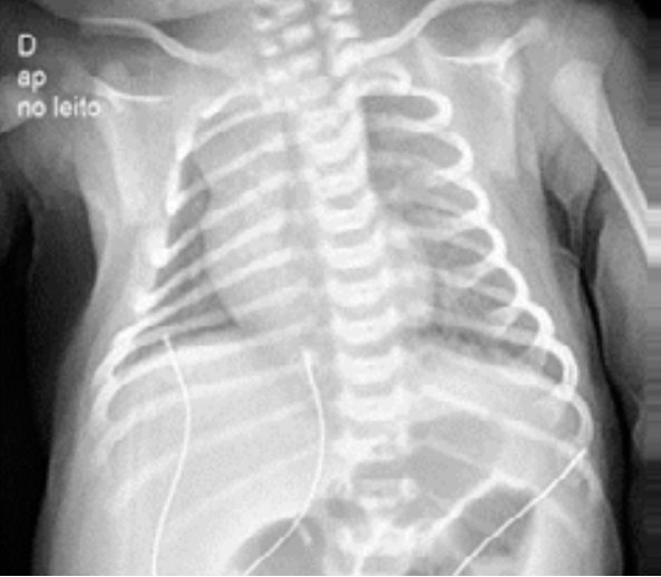
Figure 2: Chest x-ray at sixth day of life showing complete reabsorption of the pneumomediastinum and pneumothorax. 

Pneumomediastinum is uncommon in neonates (2.5:1000 live births) but should be kept in mind particularly in cases of preterm neonates, meconium aspiration, positive pressure ventilation or congenital pneumonia.1 Patients are usually asymptomatic, requiring no specific treatment, but should be monitored for evidence of cardio-respiratory compromise or development of other air leaks, especially pneumothorax. Infants with tension pneumomediastinum should be treated urgently with ultrasound-guided percutaneous drainage.

A chest x-ray should always be performed (lateral and anteroposterior views),2 although recent studies suggest a lung ultrasound can also help confirm the diagnosis.3

In the presence of a high index of suspicion, a correct and early diagnostic can prevent iatrogenic damage such as unnecessary insertion of a thoracic drain or prolonged antibiotherapy.

References

1. Steele RW, Metz JR, Bass JW, DuBois JJ. Pneumothorax and pneumomediastinum in the newborn. Radiology. 1971;98:629-32. doi: 10.1148/98.3.629. [ Links ]

2. Raissaki M, Modatsou E, Hatzidaki E. Spontaneous pneumomediastinum in A term newborn: atypical radiographic and ct appearances. BJR | Case Rep. 2019;5:20180081. [ Links ]

3. Küng E, Habrina L, Berger A, Werther T, Aichhorn L. Diagnosing pneumomediastinum in a neonate using a lung ultrasound. Lancet. 2021;398:e13. doi: 10.1016/S0140-6736(21)01592-0. [ Links ]

Responsabilidades éticas

Conflitos de interesse: Os autores declaram a inexistência de conflitos de interesse na realização do presente trabalho.

Fontes de financiamento: Não existiram fontes externas de financiamento para a realização deste artigo

Confidencialidade dos dados: Os autores declaram ter seguido os protocolos da sua instituição acerca da publicação dos dados de doentes

Consentimento: Consentimento do doente para publicação obtido

Proveniência e revisão por pares: Não comissionado; revisão externa por pares

Ethical disclosures

Conflicts of interest: The authors have no conflicts of interest to declare.

Financing support: This work has not received any contribution, grant or scholarship

Confidentiality of data: The authors declare that they have followed the protocols of their work center on the publication of data from patients

Patient consent: Consent for publication was obtained

Provenance and peer review: Not commissioned; externally peer reviewed

Received: March 14, 2022; Accepted: May 17, 2022; preprint: June 02, 2022; Published: June 30, 2022

Corresponding Author/Autor Correspondente: José Miguel Cunha de Alarcão [josecunhaalarcao@gmail.com] Rua O Conimbricense, nº66, 2ºA, 3030-504 Coimbra, Portugal ORCID iD: 0000-0002-4649-9500

JCA and RSG:

Wrote the manuscript

MN and ARS:

Critically revised the manuscript

JCA e RSG:

Escreveram o manuscrito

MN e ARS:

Fizeram a revisão crítica do manuscrito

Creative Commons License This is an open-access article distributed under the terms of the Creative Commons Attribution License