


Services on Demand
Journal
Article
 English (pdf)
 Article in xml format
 Article references
 Send this article by e-mail
Indicators
-
 Cited by SciELO
 -
 Access statistics
Related links
-
 Similars in
SciELO
Similars in
SciELO
Share

 Permalink
GE-Portuguese Journal of Gastroenterology
Print version ISSN 2341-4545
GE Port J Gastroenterol vol.26 no.6 Lisboa Dec. 2019
https://doi.org/10.1159/000495701
IMAGES IN GASTROENTEROLOGY AND HEPATOLOGY
Multiple Gastric Diverticula
Divertículos Gástricos Múltiplos
Gisela Silva, Helena Moreira Silva, Marta D. Tavares, Rosa Lima
Pediatric Gastroenterology Unity of Centro Materno-Infantil do Norte, Centro Hospitalar e Universitário do Porto, Porto, Portugal
* Corresponding author.
Keywords: Congenital diverticula, Esophageal atresia, Child
Palavras-Chave: Divertículos congénitos, Atrésia esofágica, Criança
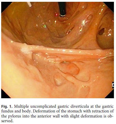
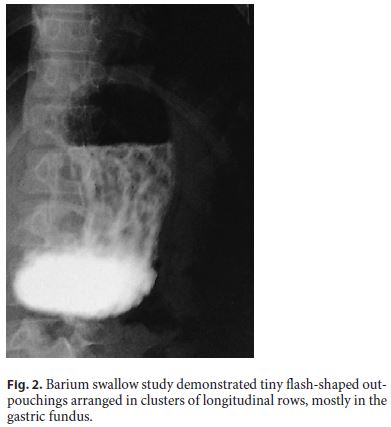
A 9-year-old girl presenting with an esophageal atresia (EA) with tracheoesophageal fistula underwent surgical correction and was submitted to examination of the esophageal structure. She presented with dyspeptic complaints. Upper gastrointestinal endoscopy (UGE) showed multiple uncomplicated gastric diverticula (GD) (Fig. 1). This pathology was confirmed with a barium swallow study (Fig. 2). Reviewing the patient’s record, we found a description of a pneumoperitoneum following gastric rupture before the correction of the EA. The esophagogram, performed at 50 days of life, showed good passage of the contrast material but irregular gastric filling and UGE revealed exaggerated folds and areas of small pockets in the lesser curvature of the stomach. GD are a rare endoscopic finding, with an estimated prevalence of 0.01–0.11% [1]. Most GD are single and acquired during life [1]. Usually, symptoms are unspecific [2]. Occasionally, patients with GD can have dramatic presentations related to massive bleeding or perforation [3]. The description of a gastric rupture in the first day of life, the localization of the GD and the association with other gastrointestinal abnormalities as EA make the diagnosis in this case more likely to be multiple congenital GD [2, 3]. This last association may be explained by embryogenesis [3]. The surgery may have contributed to the appearance of new pseudodiverticula as a consequence of perigastric adhesions in the gastric suture itself.
References
1 Palmer ED. Gastric diverticula. Int Abstr Surg. 1951 May;92(5):417–28. [ Links ]
2 Ramai D, Ofosu A, Reddy M. Gastric Diverticula: A Review and Report of Two Cases. Gastroenterol Res. 2018 Feb;11(1):68–70. [ Links ]
3 Cheng EH, Pavelock RR. Multiple gastrointestinal tract diverticula. Gastrointest Radiol. 1990;15(4):282–4. [ Links ]
Statement of Ethics
The authors have no ethical conflicts to disclose.
Disclosure Statement
The authors have no conflicts of interest to declare.
* Corresponding author.
Gisela Silva
Largo da Maternidade de Júlio Dinis
PT–4050-651 Porto (Portugal)
E-Mail giselavaqueiro@yahoo.com.br
Received: September 15, 2018; Accepted after revision: November 22, 2018

