


Services on Demand
Journal
Article
 English (pdf)
 Article in xml format
 Article references
 Send this article by e-mail
Indicators
-
 Cited by SciELO
 -
 Access statistics
Related links
-
 Similars in
SciELO
Similars in
SciELO
Share

 Permalink
GE-Portuguese Journal of Gastroenterology
Print version ISSN 2341-4545
GE Port J Gastroenterol vol.27 no.5 Lisboa Oct. 2020
https://doi.org/10.1159/505272
IMAGES IN GASTROENTEROLOGY AND HEPATOLOGY
An Unusual Small Bowel Lesion
Lesão atípica do intestino delgado
Maria Pia Costa-Santosa, João Moreira-Pintob, Maria Helena Oliveirac, Alexandre Oliveira Ferreiraa
aGastroenterology Department, Hospital Beatriz Ângelo, Loures, Portugal; bOncology Department, Hospital Beatriz Ângelo, Loures, Portugal; cPathology Department, Hospital Beatriz Ângelo, Loures, Portugal
* Corresponding author.
Keywords: Leiomyosarcoma, Small bowel, Upper gastrointestinal bleeding
Palavras-Chave: Leiomiossarcoma, Intestino delgado, Hemorragia digestiva alta
Case Report
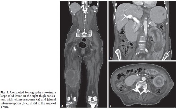
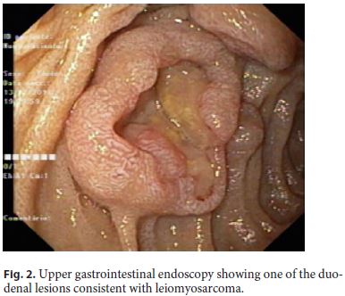
A 58-year-old black female, born in Angola and living in Portugal, presented with melena, occasional vomiting, and asthenia for 1 month. She also complained of anorexia and weight loss (40 kg/88 lb) in the past 2 years and reported a 7-year history of a right thigh mass that had progressively increased in size, with an inconclusive biopsy performed in Angola. Her laboratory tests revealed iron deficiency anemia (hemoglobin 4.4 g/dL). Computed tomography (CT) showed a large solid lesion in the right thigh (Fig. 1a), multiple smaller lesions in the thyroid, lungs, pancreas, adrenal gland, uterus, and soft tissues, focal thickness of the small bowel wall and colon, as well as jejunal intussusception, distal to the angle of Treitz (Fig. 1b, c). Upper gastrointestinal endoscopy revealed multiple ulcerative mucosal lesions with elevated edges throughout the second and third portions of the duodenum, with 10–20 mm, without hemorrhagic stigmata and surrounded by normal mucosa (Fig. 2). The duodenal and thigh lesions were biopsied, and histology showed irregularly intersecting fascicles of spindled cells with indistinct cytoplasmic contour and a cigar-shaped nucleus and areas of necrosis. Immunohistochemically, the tumor cells were positive for α-smooth muscle actin, desmin, and hcaldesmon, and negative for CD117, CK AE1/AE3, and S-100 protein. Therefore, a stage IV leiomyosarcoma was diagnosed. Due to persistent melena, endoscopy was repeated and showed an ulcerative lesion in the duodenum with active oozing. Hemostasis was achieved after adrenaline injection and coagulation with argon plasma. Nausea and vomiting relieved with conservative measures and jejunal intussusception reduced spontaneously. The patient started chemotherapy with doxorubicin. However, there was disease progression and she died 8 months after the diagnosis.
Discussion
Leiomyosarcoma is a rare smooth muscle malignant neoplasm that accounts for 10–20% of all soft tissue sarcomas [1]. It appears most commonly in the retroperitoneum, large blood vessels, and uterus, and less frequently in the lower extremities [1, 2]. Both primary and metastatic leiomyosarcoma of the small bowel are extremely rare [3]. In our case, the primary tumor is unknown since the diagnosis was made at a late stage of the disease, although clinical history strongly suggests that the lower limb might have been the origin. Gastrointestinal bleeding and intestinal obstruction are the most common presenting signs of a small bowel leiomyosarcoma [1]. Surgical resection is the treatment of choice in patients with localized disease [2]. In the present case, multiple ulcerative lesions of the small bowel caused gastrointestinal bleeding and intestinal intussusception, leading to suboclusion. However, surgery was not considered since she had disseminated disease and bowel obstruction relieved with conservative measures. In fact, intussusception often reduces spontaneously, although it can recur [3]. Small bowel metastasis by leiomyosarcoma is usually a sign of extensive disease and carries an unfavorable prognosis. Data on chemotherapy for unresectable metastatic leiomyosarcoma are scarce [2]. The most commonly used drugs as initial treatment in metastatic soft tissue sarcomas – doxorubicin and ifosfamide – show limited efficacy for leiomyosarcoma, with response rates between 10 and 25% [1, 2].
References
1 Hilal L, Barada K, Mukherji D, Temraz S, Shamseddine A. Gastrointestinal (GI) leiomyosarcoma (LMS) case series and review on diagnosis, management, and prognosis. Med Oncol. 2016 Feb;33(2):20. [ Links ]
2 Serrano C, George S. Leiomyosarcoma. Hematol Oncol Clin North Am. 2013 Oct;27(5):957–74.
3 Guzel T, Mech K, Mazurkiewicz M, Dąbrowski B, Lech G, Chaber A, et al. A very rare case of a small bowel leiomyosarcoma leading to ileocaecal intussusception treated with a laparoscopic resection: a case report and a literature review. World J Surg Oncol. 2016 Feb;14(1):48. [ Links ]
Statement of Ethics
The authors declare that this case did not require informed consent or approval by the ethics committee.
Disclosure Statement
The authors have no conflicts of interest to declare.
Funding Sources
No funding was received for this work.
* Corresponding author.
Maria Pia Costa-Santos
Gastroenterology Department, Hospital Beatriz Ângelo
Avenida Carlos Teixeira 3
PT–2674-514 Loures (Portugal)
Received: October 8, 2019; Accepted: December 6, 2019
Author Contributions
Maria Pia Costa-Santos drafted the manuscript and reviewed the literature. Joao Moreira-Pinto, Maria Helena Oliveira, and Alexandre Oliveira Ferreira contributed with critical review of the manuscript and approval of the final version.

