SciELO - Scientific Electronic Library Online

 
vol.28 número5Síndrome de ball-valve, uma apresentação incomum de GISTComentário ao artigo: Terapêutica endoscópica de vólvulo do sigmoide numa população debilitada - qual a relevância? índice de autoresíndice de assuntosPesquisa de artigos
Home Pagelista alfabética de periódicos  

Serviços Personalizados

Journal

Artigo

Indicadores

Links relacionados

  • Não possue artigos similaresSimilares em SciELO

Compartilhar


GE-Portuguese Journal of Gastroenterology

versão impressa ISSN 2341-4545versão On-line ISSN 2387-1954

GE Port J Gastroenterol vol.28 no.5 Lisboa out. 2021  Epub 20-Mar-2022

https://doi.org/10.1159/000512090 

Images in Gastroenterology and Hepatology

Anorectal Amelanotic Melanoma

Melanoma anorretal amelanótico

Akira Hokamaa 

Tetsuya Ohiraa 

Jiro Fujitab 

aDepartment of Endoscopy, Graduate School of Medicine, University of the Ryukyus, Okinawa, Japan;

bDepartment of Infectious Diseases, Respiratory and Digestive Medicine, Graduate School of Medicine, University of the Ryukyus, Okinawa, Japan


Keywords Anorectal melanoma; Rectum; Endoscopy

Palavras Chave Melanoma anorectal; Recto; Endoscopia

Case Report

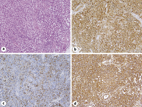
A 65-year-old man presented with a 3 months’ history of hematochezia and anal pain. Digital examination revealed a firm rectal mass. Laboratory examination revealed hemoglobin of 12.9 g/dL (normal range, 13.7-16.8 g/dL). Colonoscopy showed an anorectal tumor involving the distal 3 cm of the rectum, suggesting adenocarcinoma (Fig. 1). Endoscopic biopsies showed a round cell tumor without melanin pigmentation (Fig. 2a). Immunohistopathological examinations disclosed positive findings for S-100 (Fig. 2b), human melanin black-45 (Fig. 2c), and vimentin (Fig. 2d). A diagnosis of anorectal amelanotic melanoma was made. Computed tomography scans and positron emission tomography images disclosed multiple metastatic lesions in the lungs and liver. The patient was informed about chemotherapy options and prognosis of stage IV melanoma but refused undergoing chemotherapy. Best supportive care was offered, and the patient died 4 months later.

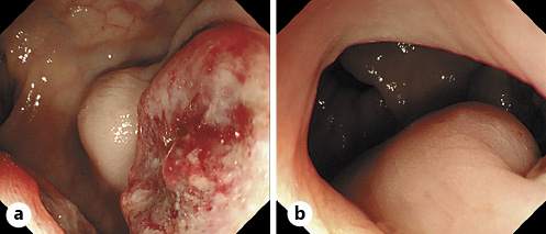
Fig. 1 Colonoscopic images of the tumor. a An ulcerated mass with submucosal invasion in the rectum. b Subepithelial invasion to the anal canal. 

Fig. 2 Histopathology of the anorectal amelanotic melanoma. a Infiltration of round cell tumor without melanin pigmentation. Hematoxylin and eosin. ×100. b Immunohistochemistry of amelanotic melanoma cells. S-100+. ×100. c Human melanin black-45+. ×100. d Vimentin+. ×100. 

Discussion

Anorectal amelanotic melanoma is an extremely rare disease [1]. As amelanotic melanoma has few melanin granules, it is not easy to differentiate it from various anorectal tumors, such as adenocarcinoma, squamous cell carcinoma, lymphoma, and gastrointestinal stromal tumor. Therefore, immunostaining, as shown in this case, is the key for the definite diagnosis [1, 2]. The prognosis of anorectal melanoma remains poor, as most cases have lymph node or distal metastasis at the time of diagnosis. Although surgery remains the optimal treatment option, it is regarded as a palliative procedure, making no significant improvement in overall survival [3]. Conventionally, cytotoxic agents, such as dacarbazine and cisplatin, have been applied to stage IV melanoma, with little improvement of prognosis [2, 3]. Recent development of immunotherapy including immune-check point inhibitors, such as nivolumab and pembrolizumab, have improved the prognosis of this highly lethal malignancy [2, 3]

References

1 Hillenbrand A, Barth TF, Henne-Bruns D, Formentini A. Anorectal amelanotic melanoma. Colorectal Dis. 2008 Jul;10(6):612-5. [ Links ]

2 Tokuhara K, Nakatani K, Tanimura H, Yoshioka K, Kiyohara T, Kon M. A first reported case of metastatic anorectal amelanotic melanoma with a marked response to anti-PD-1 antibody nivolumab: a case report. Int J Surg Case Rep. 2017;31:188-92. [ Links ]

3 Taylor JP, Stem M, Yu D, Chen SY, Fang SH, Gearhart SL, et al. Treatment strategies and survival trends for anorectal melanoma: is it time for a change? World J Surg. 2019 Jul;43(7):1809-19. [ Links ]

Statement of Ethics Consent from the patient was obtained for publication. This paper was conducted ethically in accordance with the Declaration of Helsinki

Funding Sources The authors have no funding sources to declare

Received: August 12, 2020; Accepted: September 28, 2020

Corresponding author Akira Hokama Department of Endoscopy Graduate School of Medicine, University of the Ryukyus 207 Uehara, Nishihara, Okinawa 903-0215 (Japan) hokama-a@med.u-ryukyu.ac.jp

Conflict of Interest Statement

The authors have no conflicts of interest to declare

Author Contributions

A.H. performed the endoscopy, obtained the pathological images, and drafted the manuscript. T.O. and J.F. reviewed the paper and approved the final version

Creative Commons License This is an open-access article distributed under the terms of the Creative Commons Attribution License