SciELO - Scientific Electronic Library Online

 
vol.28 número6Pneumatose cística intestinal: uma causa rara de hemorragia gastrointestinalErratum índice de autoresíndice de assuntosPesquisa de artigos
Home Pagelista alfabética de periódicos  

Serviços Personalizados

Journal

Artigo

Indicadores

Links relacionados

  • Não possue artigos similaresSimilares em SciELO

Compartilhar


GE-Portuguese Journal of Gastroenterology

versão impressa ISSN 2341-4545versão On-line ISSN 2387-1954

GE Port J Gastroenterol vol.28 no.6 Lisboa dez. 2021  Epub 30-Mar-2022

https://doi.org/10.1159/000514008 

Images in Gastroenterology and Hepatology

A Rare Cause of Cholestasis Related to Polycystic Liver Disease

Uma causa rara de colestase relacionada com doença hepática poliquística

Pedro Costa-Moreiraa  b 

Susana Lopesa  b 

Guilherme Macedoa  b 

aGastroenterology Department, Centro Hospitalar Universitário São João, Porto, Portugal;

bFaculty of Medicine, University of Porto, Porto, Portugal


Keywords Polycystic liver disease; Vascular liver disease; Cholestasis

Palavras Chave Doença hepática poliquística; Doença vascular hepática; Colestase

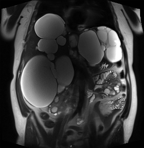
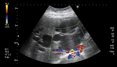
A 52-year-old female with autosomal dominant polycystic kidney disease (ADPKD) associated with polycystic liver disease (PLD) was referred to the gastroenterology clinic due to abnormal liver function tests. Laboratory testing demonstrated increased γ-glutamyltransferase (1,231 vs. normal <40 IU/L) and alkaline phosphatase (218 vs. normal <120 IU/L) levels. The patient was asymptomatic, and there were no clinical manifestations of portal hypertension. The etiological study for liver disease (including viral serologies, autoimmunity, iron kinetics, and ceruloplasmin) was negative. Abdominal magnetic resonance imaging (MRI) showed multiple hepatic cysts (the largest with a 13-cm diameter) (Fig. 1). Abdominal ultrasound with Doppler imaging showed a normal patency of the portal and hepatic veins (Fig. 2). A parenchymal liver biopsy showed marked centrilobular congestion with blood-filled cavities that resembled hepatic peliosis and was associated with findings of mild portal and intralobular inflammation (Fig. 3, 4).

Fig. 1: MRI of large hepatic cysts distributed in the liver parenchyma. 

Fig. 2: Abdominal ultrasound with Doppler imaging of the portal vein. HE. ×100. 

Fig. 3: Liver biopsy showing centrilobular congestion. HE. ×200. 

Fig. 4: Liver biopsy showing centrilobular congestion. 

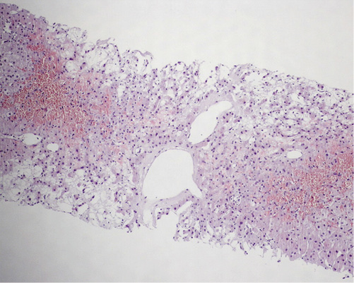
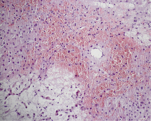
After the diagnostic workup, we concluded that the patient had hepatic venous outflow obstruction secondary to cystic vascular compression. While the liver architecture is affected by PLD, the synthetic function of the polycystic liver remains intact until very late in the course of the disease [1, 2]. Histological features of this functional disease include centrizonal congestion, necrosis, and hemorrhage. Large regenerative nodules, obstructive portal venopathy, and fibrosis/cirrhosis may also be found [3].

It is important to note that in the classical forms of peliosis hepatis, the lesions are randomly distributed in the liver parenchyma. However, in the case of vascular outflow obstruction, the sinusoidal dilation is more evident in the centrilobular area (zone 3) [4].

This case highlights a rare vascular liver disease related to a genetic condition that wouldn’t usually have a significant impact on liver function. Early diagnosis may be helpful, allowing for a strategic cyst intervention that can avoid the consequences of portal hypertension.

References

1 DeLeve LD, Valla DC, Garcia-Tsao G; American Association for the Study Liver Diseases. Vascular disorders of the liver. Hepatology. 2009 May;49(5):1729-64. [ Links ]

2 van Aerts RM, van de Laarschot LF, Banales JM, Drenth JP. Clinical management of polycystic liver disease. J Hepatol. 2018 Apr;68(4):827-37. [ Links ]

3 Bernts LH, Drenth JP, Tjwa ET. Management of portal hypertension and ascites in polycystic liver disease. Liver Int. 2019 Nov;39(11):2024-33. [ Links ]

4 Uddin W, Ramage JK, Portmann B, Wilson P, Benjamin I, Tan KC, et al. Hepatic venous outflow obstruction in patients with polycystic liver disease: pathogenesis and treatment. Gut. 1995 Jan;36(1):142-5. [ Links ]

Statement of Ethics The authors confirm that written informed consent was obtained for the publication of this case (including the images)

Funding Sources There was no funding

Received: October 13, 2020; Accepted: December 04, 2020

Corresponding author Pedro Manuel Costa Moreira Gastroenterology Department Centro Hospitalar Universitário São João Al. Prof. Hernâni Monteiro PT-4200-319 Porto (Portugal) pedromoreira.med@gmail.com

Conflict of Interest Statement

All authors disclosed that there are no personal conflicts of interest or financial relationships relevant to this publication

Author Contributions

P.C.-M.: manuscript concept and design, literature review, and draft of the manuscript. S.L. and G.M.: critical revision of the manuscript. All authors approved the final version

Creative Commons License This is an open-access article distributed under the terms of the Creative Commons Attribution License