


 Inglês (pdf)
 Artigo em XML
 Referências do artigo
 Enviar este artigo por email
 Citado por SciELO
  Similares em
SciELO
Similares em
SciELO 
 Permalink
An 84-year-old female was referred for exclusion of gastrointestinal bleeding for anemia workup (hemoglobin 8.1 g/dL; mean corpuscular volume 86 fL). With a view to clinical, laboratory and endoscopic findings, the latter was ex post considered to represent anemia of chronic disease with a putative renal component. Medical history included curatively treated breast cancer, diabetic chronic kidney disease (Dialysis Outcome Quality Initiative stage 3A) and rheumatoid arthritis. The patient was on multiple chronic medications including hydrochloroquine, cholecalciferol, torasemide, letrozole, iron-(II)-sulfate and a mixture of dissolved and isophane insulin. Of note, there was no recent intake of any nonsteroidal anti-inflammatory drugs.
The recent routine esophagogastroduodenoscopy was significant for a duodenal mucosa diffusely speckled by multiple tiny brownish-black spots (Fig. 1a). Image-enhanced magnification endoscopy of the duodenal surface indicated a well-preserved villous architecture with linked color imaging (Eluxeo, Fuji, Düsseldorf, Germany) highlighting brownish pigment in the villous tips (1) (Fig. 1b). Histopathology likewise demonstrated brown pigmented granules within macrophages with positive iron staining studies, replicating the endoscopic diagnosis of pseudomelanosis duodeni (Fig. 1c). An ancillary small-bowel capsule endoscopy (PillCam SB3, Medtronic, Meerbusch, Germany) could reproduce these findings to be limited to the duodenal mucosal lining, and, in concert with an unrevealing ileocolonoscopy, a potential gastrointestinal bleeding source was firmly excluded (Fig. 1d).
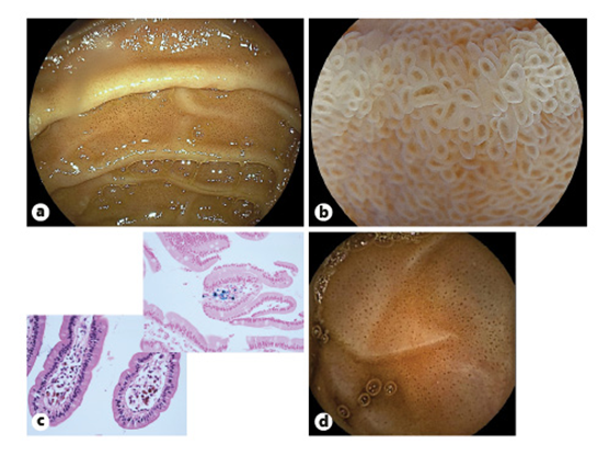
Fig. 1: a Routine esophagogastroduodenoscopy demonstrating a duodenal mucosa speckled by multiple tiny brownish to black spots in the descending duodenum. b Cap-fitted image-enhanced magnification endoscopy using linked color imaging indicated an intact villous architecture with brownish deposits in the villous tips. c Histopathology of duodenal biopsies indicated pigment within macrophages in the tip of the villi with iron stains confirming iron deposits (left: hematoxylin-eosin, ×20; right: Prussian blue, ×20). d Small-bowel capsule endoscopy replicated these findings and confirmed limitation to the duodenum, while excluding a potential small-bowel bleeding source.
Pseudomelanosis duodeni is an incidental, yet engrammatic endoscopic finding with no known clinical relevance (2). Albeit its distinct pathogenesis has to be better delineated, clinical associations exist as to chronic kidney disease and altered duodenal iron transport and sulfa moieties, included in certain mostly antihypertensive medications typically linked to it, such as hydralazine (3). As is illustrated here, specialized high-quality endoscopy is instrumental in rapid identification and characterization of its engrammatic endoscopic features as well as forestalling the characteristic underlying histopathology.

