


 Inglês (pdf)
 Artigo em XML
 Referências do artigo
 Enviar este artigo por email
 Citado por SciELO
  Similares em
SciELO
Similares em
SciELO 
 Permalink
A 29-year-old woman presented with a voluminous hepatic mass on a screening ultrasound performed during her second pregnancy. She underwent a magnetic resonance imaging scan that was consistent with a cavernous hemangioma measuring 250 mm in the largest diameter, with a nodular margin, hypointense images on T1-weighted sequences, hyperintense on T2-weighted sequences, and higher signal intensity on long repetition and echo times. This mass involved almost the entire right hepatic lobe with multiple infracentimetric hemangiomas in the left lobe. An analytical study showed discrete pancytopenia. Liver tests were within the normal range. Both her pregnancies were uneventful. Due to the absence of symptoms the patient was kept under clinical surveillance.
During 25 years of follow-up the patient remained asymptomatic, with a progressive increase of lesion dimensions. A subsequent computed tomography scan revealed a hypodense mass with centrifugal enhancement during the late phase, an atypical presentation in relation to its dimensions (351 × 180 × 220 mm; Fig. 1).
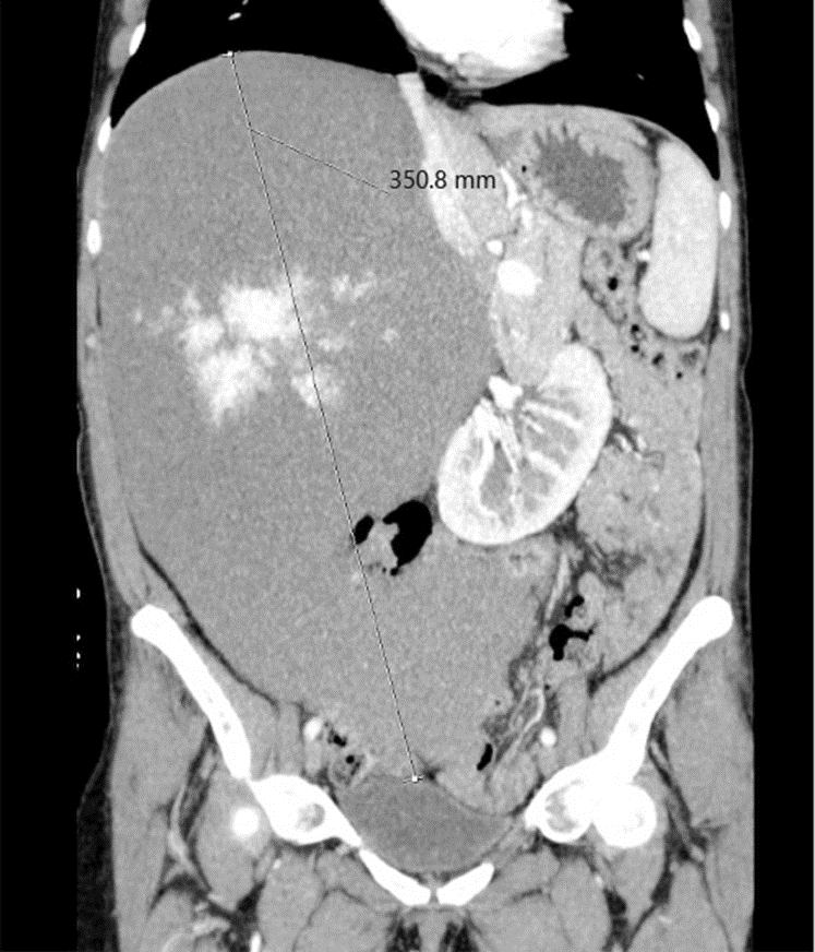
Fig. 1: Contrast-enhanced coronal computed tomography scan demonstrating the huge size of the hemangioma (351 × 180 × 220 mm), which occupies almost the entire right lobe.
This massive liver lesion in the right lobe lead to considerable displacement of the pancreas, descending colon, small bowel, and the right kidney to the opposite side (Fig. 2). There were no signs of hepatic venous outflow obstruction, portal hypertension, or compression of the biliary tract.
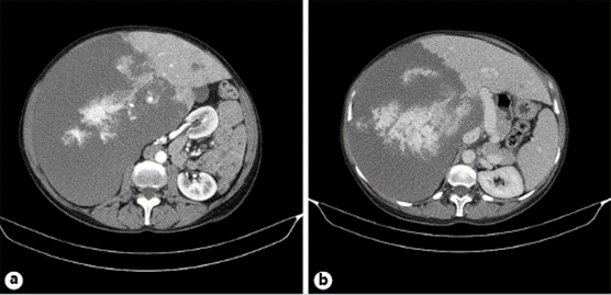
Fig. 2: Contrast-enhanced axial computed tomography scan showing the displacement of the pancreas, descending colon, small bowel, and the right kidney to the left side, due to the large size of the hemangioma in the right hepatic lobe. a Arterial phase. b Portal venous phase.
When hemangiomas exceed 50 mm, they are defined as giant lesions (1). Here, we present a ”super-giant” hemangioma, diagnosed during the second pregnancy of the patient, showing a slow growth rate and no complications after more than 2 decades of follow-up.
The influence of gender in liver hemangiomas appearance is undeniable according to a female:male ratio of up to 5:1 (2). However, the role of female sex hormones and the effect of pregnancy on the evolution of hemangiomas is uncertain. There are reports of hemangioma complications during pregnancy such as rupture and intratumoral hemorrhage (3, 4), but growth of hepatic hemangiomas during pregnancy is difficult to estimate due to the lack of previous imaging evaluations. Giant hemangiomas should not preclude pregnancy, but close monitoring should be provided.

