


 Inglês (pdf)
 Artigo em XML
 Referências do artigo
 Enviar este artigo por email
 Citado por SciELO
  Similares em
SciELO
Similares em
SciELO 
 Permalink
A Caucasian 60-year-old man, with a career in house-building, presented with mild, chronic and progressive dysphagia, manifested by difficulty in swallowing solid food for the last 6 months. He had no cervical pain, weight loss, dysphonia, respiratory complaints, previous surgical intervention, radiotherapy, major trauma, myogenic or metabolic known causes for this symptom. The patient had a normal cervical and chest physical examination and neurologic exam. A barium swallow test was firstly performed, revealing a posterior esophagus indentation due to an osteophyte formation, which interfered with contrast progression. This exam also ruled out dysfunction in motility or a Zenker’s diverticulum. Consequently, to better characterize the osteoarticular disease, the patient was submitted to a computed tomography (CT) and cervical magnetic resonance imaging (MRI) which revealed degenerative changes such as: anterior and lateral protrusion of vertebral discus, osteophytes from C3 to C5, uncarthrosis, and posterior C3 to C6 interapophysary hypertrophic arthrosis, without ligament calcification or cervical webs (Fig. 1a-c). Finally, upper digestive endoscopy ruled out intrinsic abnormalities, and cervical ultrasound was also normal.
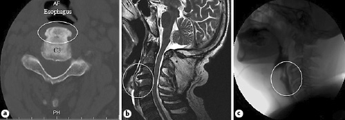
Fig. 1 a C3 somatic marginal osteophyte (white circle) on CT. b Cervical MRI with anterior and lateral protrusion of the vertebral discus and osteophytes in C3 to C5. c Barium swallows with posterior esophagus indentation at the level of the osteophyte formation.
Cervical osteophytes occur in 20-30% of the general population [1] and are mostly associated with diffuse idiopathic skeletal hyperostosis and ankylosing spondylitis [2]. Most osteophytes of the anterior margin of the cervical spine are asymptomatic. Nevertheless, cervical osteophytes were the cause of dysphagia in 11.7% of a studied population, with a mean age of 79 ± 8 years [3, 4], disclosing a possible underrecognized etiology. In this matter, according to Lee et al. [5], patients with this condition were usually men (81%), with a mean age of 68 years and the C3 to C6 level most commonly involved, typically presenting with a long history of dysphagia. Other symptoms include food impaction, dysphonia and respiratory obstruction [2, 3]. The diagnostic imaging approach includes a lateral X-ray, barium swallow, and cervical CT or MRI. Surgical approach may be a definite solution, but diet restrictions may cause satisfactory improvement [1-3, 5]. Our patient was paucisymptomatic and remains only with adapted diet. The authors highlight anterior cervical osteophytosis as a differential diagnosis of oropharyngeal dysphagia.

