SciELO - Scientific Electronic Library Online

 
vol.29 número6Heterotopia gástrica: uma causa rara de impactação alimentar índice de autoresíndice de assuntosPesquisa de artigos
Home Pagelista alfabética de periódicos  

Serviços Personalizados

Journal

Artigo

Indicadores

Links relacionados

  • Não possue artigos similaresSimilares em SciELO

Compartilhar


GE-Portuguese Journal of Gastroenterology

versão impressa ISSN 2341-4545

GE Port J Gastroenterol vol.29 no.6 Lisboa dez. 2022  Epub 02-Jan-2023

https://doi.org/10.1159/000518446 

Images in Gastroenterology and Hepatology

Ultrasound Trilaminar Sign of Abdominal Cocoon

Sinal trilaminar ecográfico do casulo abdominal

Venkatraman Indiran1 

Dillibabu Ethiraj2 

1Department of Radio-diagnosis, Sree Balaji Medical College and Hospital, Chennai, India;

2Department of Radiodiagnosis, Bhaarath Medical College and Hospital, Chennai, India


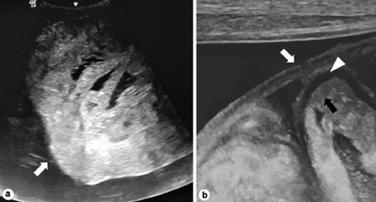
A 50-year-old male presented with vomiting and abdominal pain for 3 days. He is a known case of chronic renal failure for the past 2 years undergoing repeated peritoneal dialysis. On physical examination, his abdomen was distended. Laboratory investigation showed increased serum creatinine levels ∼6 mg/dL. Ultrasound abdomen showed shrunken kidneys with increased cortical echoes and ascites. Apart from that, thickened peritoneum or a membrane overlying the bowel loops was seen on high-frequency ultrasound as a triple-layer appearance, which is called “ultrasound trilaminar sign” (Fig. 1a, b) [1]. The “ultrasound trilaminar sign” is considered characteristic of the abdominal cocoon or encapsulating peritoneal sclerosis (EPS) [2]. EPS is characterized by clustered small bowel loops with narrow base and surrounding thick fibro-collagenous membrane. It may be idiopathic or secondary to repeated peritoneal dialysis, abdominal tuberculosis, abdominal surgeries, and drugs like propranolol [1, 3]. The three layers forming the ultrasound trilaminar appearance are the superficial hyperechoic peritoneal membrane, a middle hypoechoic layer of the bowel wall, and the deep hyperechoic layer produced by the bowel gas or contents [1]. This trilaminar appearance is not to be confused with computed tomography trilaminar sign, which represents submucosal bowel edema [1]. Presence of ascites is usually essential for identifying the trilaminar appearance and cauliflower-like appearance of clustered bowel loops. The patient was advised surgical evaluation for removal of membrane and adhesiolysis.

Fig. 1 a Ultrasound abdomen shows clumped bowel loops (white arrow) and ascites. b Ultrasound trilaminar sign of abdominal cocoon hyperechoic membrane (white arrow), hypoechoic bowel wall (arrowhead), and hyperechoic bowel contents (black arrow) 

.

References

1. Singhal M, Krishna S, Lal A, Narayanasamy S, Bal A, Yadav TD, et al. Encapsulating Peritoneal Sclerosis: The Abdominal Cocoon. Radiographics. 2019 JanFeb;39(1):62-77. [ Links ]

2. Campbell S, Clarke P, Hawley C, Wigan M, Kerlin P, Butler J, et al. Sclerosing peritonitis: identification of diagnostic, clinical, and radiological features. Am J Kidney Dis. 1994 Nov;24(5):819-25. [ Links ]

3. Ethiraj D, Indiran V. V Indiran - Abdominal Cocoon: "Cauliflower Sign" on Contrast-Enhanced Computed Tomography Scan. GE Port J Gastroenterol. 2021;28(1):76-8. [ Links ]

1Statement of Ethics All procedures performed in studies involving human participants were in accordance with the ethical standards of the institutional and/or national research committee and with the 1964 Helsinki Declaration and its later amendments or comparable ethical standards.

Funding Sources There is no funding.

Received: April 21, 2021; Accepted: July 05, 2021

Corresponding author Dillibabu Ethiraj dillibaburaghavan1993@gmail.com

Conflict of Interest Statement The authors declare that they have no conflict of interest. Informed consent was obtained from the individual participant included in the study.

Author Contributions Substantial contributions to the conception or design of the work; or the acquisition, analysis, or interpretation of data for the work - D.E., V.I. Drafting the work or revising it critically for important intellectual content - D.E., V.I. Final approval of the version to be published - D.E., V.I. Agreement to be accountable for all aspects of the work in ensuring that questions related to the accuracy or integrity of any part of the work are appropriately investigated and resolved - D.E., V.I

Creative Commons License This is an open-access article distributed under the terms of the Creative Commons Attribution License