SciELO - Scientific Electronic Library Online

 
vol.30 número4Manifestação inaugural de seminoma testicular como hemorragia digestiva: uma causa rara de metastização gástricaRemoção difícil de balão intragástrico: uma técnica diferente índice de autoresíndice de assuntosPesquisa de artigos
Home Pagelista alfabética de periódicos  

Serviços Personalizados

Journal

Artigo

Indicadores

Links relacionados

  • Não possue artigos similaresSimilares em SciELO

Compartilhar


GE-Portuguese Journal of Gastroenterology

versão impressa ISSN 2341-4545

GE Port J Gastroenterol vol.30 no.4 Lisboa ago. 2023  Epub 01-Nov-2023

https://doi.org/10.1159/000523774 

Endoscopic Snapshot

Two Cancers in One Barrett’s Segment: First Report of Concurrent Squamous Cell Carcinoma and Adenocarcinoma

Duas neoplasias num segmento de Barrett: primeiro relato de carcinoma espinocelular e adenocarcinoma concomitantes

Vincent Zimmer1  2 

Bert Bier3 

Myriam Metzger4 

Matthias Glanemann5 

1Department of Medicine, Marienhausklinik St. Josef Kohlhof, Neunkirchen, Germany;

2Department of Medicine II, Saarland University Medical Center, Saarland University, Homburg, Germany;

3Insitute of Pathology Saarbrücken-Rastpfuhl, Saarbrücken, Germany;

4Institute of Pathology, Saarland University Medical Center, Saarland University, Homburg, Germany;

5Department of General, Visceral, Vascular, and Pediatric Surgery, Saarland University Medical Center, Saarland University, Homburg, Germany


A 70-year-old male patient underwent his first upper endoscopy for anemia work-up. Beyond a 3-cm hiatal hernia, an endoscopic diagnosis of Barrett’s esophagus (Prague classification C3M4) was entertained, as illustrated by a tongue at 3 o’clock on blue laser imaging (Fig. 1a). However, at the gastroesophageal junction an estimated 20-mm nodular, superficially ulcerated lesion emerged (Fig. 1b), likewise visualized on linked color imaging (LCI) after intra-hernial retroflexion (Fig. 1c). Giv en this presumed malignant complication, full-scale assessment of the Barrett’s esophagus was warranted, including acetic acid spraying. In combination with LCI, another 8-mm utterly flat lesion emerged at 6 o’clock with an irregular vessel and surface pattern, highly suggestive of early cancer as a second lesion (Fig. 1d). Pathology of endoscopic biopsies confirmed specialized intestinal metaplasia and, more intriguingly, indicated a well-differentiated adenocarcinoma (AC) for the flat lesion (Fig. 2a) and a poorly differentiated squamous cell carcinoma (SCC) for the nodular lesion (Fig. 2b). Cross-sectional and EUS staging indicated T1/2N+ stage. Notwith-standing, due to advanced chronic obstructive pulmonary disease (GOLD IIIB with long-term oxygen therapy), the patient underwent upfront esophagectomy without significant complications after pulmonary prehabilitation. Final surgical pathology indicated pT1a, pN0(0/27), G1 for the AC and pT1b, pN1(2/27), G3 for the SCC (furthermore: L0, V0, Pn0, R0 each).
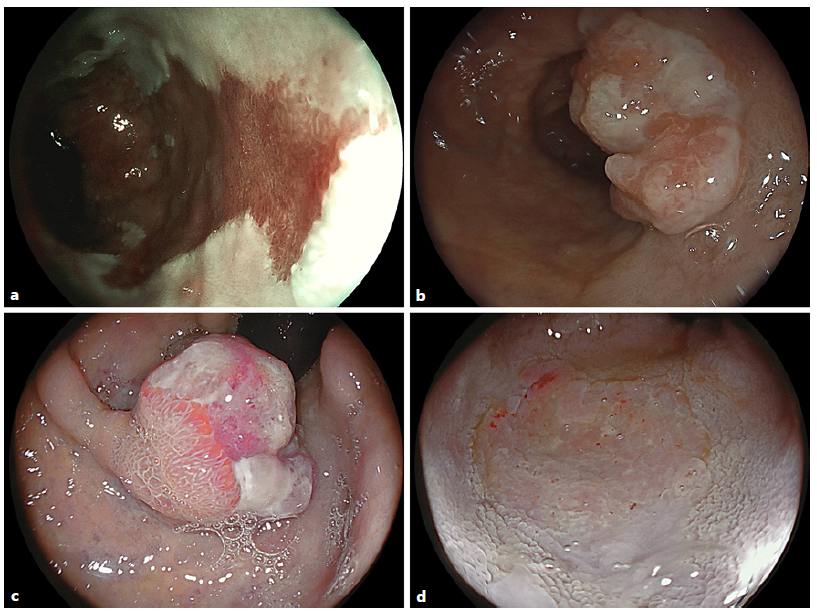
Fig. 1 a Blue laser imaging of a Prague C3M4 Barrett’s esophagus with a tongue highlighted at 3 o’clock. b An estimated 20-mm nodular ulcerated lesion emerged at the gastroesophageal junction (c) as replicated on retroflexed LCI visualization. d LCI after acetic acid spraying in the distal esophagus highlighted another 8-mm flat lesion with an irregular vessel and surface pattern consistent with early cancer. 
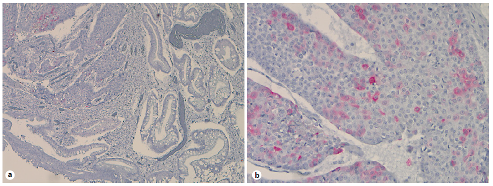
Fig. 2 Immunohistochemistries for cytokeratin (CK) 5/6 with negative staining of the AC (a) and positive results in the SCC (b). 

Barrett´s esophagus is a well-acknowledged risk factor for esophageal AC formation; however, singular cases of SCC arising in Barrett’s esophagus and/or collision tumor comprising SCC and AC elements have been documented in the literature, pointing to ambivalent carcinogenic field effects [1-3]. Concurrent SCC and Barrett’s carcinoma has occasionally been reported in Barrett’s esophagus before, however, to the best of our knowledge, not with the Barrett’s segment itself [4-6]. Of interest, the patient had a mixed risk profile, including obesity and metabolic syndrome (Barrett’s) and heavy smoking (SCC). The distinct molecular mechanisms for a presumed field cancerization within a Barrett’s esophagus, which has been discussed in the literature, however, remain elusive.

References

1. Streppel MM, Siersema PD, de Leng WW, Morsink FH, Vleggaar FP, Maitra A, et al. Squamous cell carcinoma in Barrett’s esophagus: field effect versus metastasis. Dis Esophagus. 2012; 25(7): 630-7. [ Links ]

2. Takeuchi A, Hatta W, Koike T, Saito M, Jin X, Asanuma K, et al. A primary Barrett’s adenocarcinoma with a squamous cell carcinoma component. Intern Med. 2019; 58(17): 2467-72. [ Links ]

3. Mishima Y, Amano Y, Yuki T, Kusunoki R, Oka A, Uno G, et al. A rare case of Barrett’s adenocarcinoma including squamous cell carcinoma component. Clin J Gastroenterol. 2011; 4(1): 5-9. [ Links ]

4. Yamazaki T, Iwaya Y, Iwaya M, Watanabe T, Seki A, Ochi Y, et al. A case of simultaneous esophageal squamous cell carcinoma and Barrett’s adenocarcinoma. Clin J Gastroenterol. 2016; 9(4): 222-7. [ Links ]

5. Kobayashi T, Shiozaki A, Fujiwara H, Konishi H, Arita T, Kosuga T, et al. A case of synchronous multiple esophageal cancers composed of squamous cell carcinoma and Barrett’s adenocarcinoma. Gan To Kagaku Ryoho. 2015; 42(12): 1890-2. [ Links ]

6. Maleki I, Shekarriz R, Nosrati A, Orang E. Simultaneous esophageal squamous cell carcinoma and adenocarcinoma: a case report. Middle East J Dig Dis. 2015; 7(4): 257-60 [ Links ]

1Statement of Ethics The patient gave written informed consent for publication (including the publication of images).

3Funding Sources No funding was involved in this work.

Received: September 29, 2021; Accepted: December 29, 2021

Correspondence to: Vincent Zimmer, vincent.zimmer@gmx.de

Conflict of Interest Statement The authors have no conflicts of interest to declare.

Author Contributions V.Z. - clinical care, drafting and finalization of manuscript

Creative Commons License This is an open-access article distributed under the terms of the Creative Commons Attribution License