SciELO - Scientific Electronic Library Online

 
vol.30 suppl.1Ecoendoscopia Terapêutica: Indicações Atuais e Perspetivas FuturasPróteses esofágicas: uma abordagem pessoal índice de autoresíndice de assuntosPesquisa de artigos
Home Pagelista alfabética de periódicos  

Serviços Personalizados

Journal

Artigo

Indicadores

Links relacionados

  • Não possue artigos similaresSimilares em SciELO

Compartilhar


GE-Portuguese Journal of Gastroenterology

versão impressa ISSN 2341-4545

GE Port J Gastroenterol vol.30  supl.1 Lisboa ago. 2023  Epub 15-Nov-2023

https://doi.org/10.1159/000527202 

Review Article

Best Practices in Esophageal, Gastroduodenal, and Colonic Stenting

Melhores Práticas Em Próteses Endoscópicas Esofágicas, Gastroduodenais e Colorretais

Renato Medas1  2 

Joel Ferreira-Silva1  2 
http://orcid.org/0000-0002-0224-8451

Mohit Girotra3 

Monique Barakat4 

James H. Tabibian5  6 
http://orcid.org/0000-0001-9104-1702

Eduardo Rodrigues-Pinto1  2 

1Gastroenterology Department, Centro Hospitalar São João, Porto, Portugal;

2Faculty of Medicine of the University of Porto, Porto, Portugal;

3Digestive Health Institute, Swedish Medical Center, Seattle, WA, USA;

4Division of Gastroenterology, Stanford University, Stanford, CA, USA;

5Division of Gastroenterology, Department of Medicine, Olive View-UCLA Medical Center, Sylmar, CA, USA;

6UCLA Vatche and Tamar Manoukian Division of Digestive Diseases, David Geffen School of Medicine at UCLA, Los Angeles, CA, USA


Abstract

Endoscopic stenting is an area of endoscopy that has wit-nessed noteworthy advancements over the last decade, resulting in evolving clinical practices among gastroenterologists around the world. Indications for endoscopic stenting have progressively expanded, becoming a frequent part of the management algorithm for various benign and malignant conditions of the gastrointestinal tract, from esophagus to rectum. In addition to expanded indications, continuous technological enhancements and development of novel endoscopic stents have resulted in an increased success of these approaches and, in some cases, allowed new applications. This review aimed to summarize best practices in esophageal, gastroduodenal, and colonic stenting.

Keywords: Endoscopic stenting; Esophageal obstruction; Gastric outlet obstruction; Colonic obstruction

Resumo

A colocação de próteses endoscópicas é uma técnica que tem testemunhado avanços notáveis na última década, resultando na evolução da prática clínica diária dos gastroenterologistas em todo o mundo. As indicações para a colocação de próteses endoscópicas têm expandido progressivamente, tornando-se uma opção cada vez mais frequente no algoritmo de abordagem das mais variadas condições benignas e malignas do trato gastrointestinal (desde o esófago ao reto). Além da expansão nas indicações, o aprimoramento tecnológico contínuo e o desenvolvimento de novas próteses endoscópicos resultaram num maior sucesso dessas abordagens e, em alguns casos, permitiram novas aplicações. Esta revisão tem como objetivo resumir as melhores práticas em colocação de próteses endoscópicas esofágicas, gastroduodenais e colorretais.

Palavras Chave: Próteses endoscópicas; Estenose esofágica; Obstrução de saída gástrica; Estenose cólica

Introduction

Over the last few years, therapeutic endoscopy has evolved into the preferred approach, or at least a valid alternative, for management of several gastrointestinal (GI) conditions, wherein surgery was considered the standard therapy for decades [1]. Endoscopic stenting is one such aspect of therapeutic endoscopy that has witnessed noteworthy advancements. Traditionally, the main indications for endoscopic stenting were limited to the palliation of malignant disorders, such as obstructive esophageal cancer, malignant gastric outlet obstruction (GOO), and malignant colonic obstruction [2]. More recently, indications for endoscopic stenting have gradually expanded to include a variety of nonmalignant/non-obstructive disorders, such as external compression of the GI tract, GI transmural defects (e.g., perforations, fistulae, and leaks), and selected cases of refractory benign strictures [3]. Moreover, advances in biotechnology and clinical expertise have helped mitigate stent-related adverse events (AEs) [4]. In this review, we aim to summarize the evidence and experience supporting the best practices in luminal endoscopic stenting, with a specific focus on esophageal, gastroduodenal, and colonic stenting (Table 1).

Table 1 Summary of best evidence for each indication regarding different types of stents 

Esophageal Stenting

Malignant Esophageal Cancer

Palliation of Malignant Dysphagia

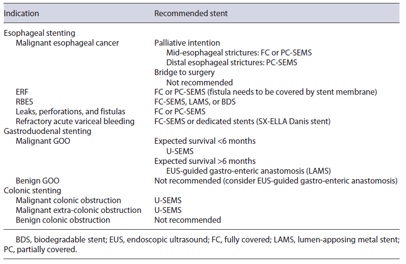
The main goal of esophageal stenting is palliation of malignant dysphagia in patients with esophageal cancer to improve nutritional intake. Although stenting provides a rapid relief of dysphagia symptoms, it is preferable in patients with an expected short survival (<3 months) (Fig. 1) [5]. A meta-analysis by Wang et al. included three randomized clinical trials (RCTs) and showed similar outcomes between fully covered self-expandable metal stent (FC-SEMS) and partially covered SEMS (PC-SEMS), without differences in stent migration, obstruction, or bleeding [6]. Pooled data from available studies showed a major AE rate of 18% with PC-SEMS and 21% with FC-SEMS (most frequently reflux, severe pain, bleeding, and ingrowth/overgrowth) [7]. Different stent designs have been developed in order to prolong stent patency and reduce AEs; however, this is hard to accomplish as stents do not affect natural history of the disease. Regarding anti-reflux stents, for example, a 2019 meta-analysis [8] and a subsequent RCT [9] failed to prove their superiority regarding improvement of reflux, dysphagia score, or related AEs (stent migration, bleeding, and obstruction).

Fig. 1 Patient with dysphagia secondary to an esophageal squamous cell carcinoma located in the mid esophagus. a Endoscopic image showing proximal view of the lesion. b, c Fluoroscopic and endoscopic images after placement of a partially covered 150 × 20 mm self-expandable metal stent. 

One major drawback of stent use in patients with longer survival is the increased risk of stent dysfunction and AE occurrence. Even though SEMSs are associated with earlier symptom relief, for patients with longer expected survival (≥3 months), brachytherapy seems to provide better quality of life, long-term dysphagia relief, and fewer AEs, when compared to SEMS placement [10, 11]. However, despite being associated with better long-term results, brachytherapy is underused in clinical practice [12, 13]. Overall severe AEs from brachytherapy alone may occur in up to 23% of cases, mostly including brachy-therapy-related stenosis (12%) and fistula formation (8%) [14]. The effect of combined brachytherapy and stenting on AE rates is not completely clear; however, it seems to provide better dysphagia relief in patients with survival longer than 3 months and higher overall survival, compared to SEMS alone [15]. Patients may also be palliated with external beam radiotherapy (EBRT) alone [16, 17]. A recent propensity score-matched analysis that compared EBRT alone with brachytherapy alone suggested that EBRT may offer a faster and safer dysphagia relief compared to brachytherapy, with similar long-term outcomes [18]. A recent RCT that compared EBRT alone with a combination of EBRT and chemotherapy found that EBRT alone had similar dysphagia relief and survival as the combination therapy, but fewer AEs [19].

Some retrospective cohorts evaluated patients submitted to SEMS placement, with ≥6-month survival, and concluded that SEMS may be a valid alternative, especially in centers where brachytherapy is not widely available. Despite the increased risk of AEs over time, most of them can be managed endoscopically [20-22].

Irradiation stents have been developed to combine advantages of both SEMS and radiotherapy. A 2017 [23] and a 2021 [24] meta-analysis comparing irradiation SEMS (loaded with 125I beads) versus traditional SEMS showed prolonged patient survival and stent patency with irradiation stents, with no differences in AE rates. Biodegradable stent (BDS) role in the palliation of malignant dysphagia is not adequately defined and should not yet be considered a valid alternative to SEMS [25].

Recommendation: Patients with life expectancy of less than 3 months or suffering from severe dysphagia should be considered for SEMS placement. FC- or PC-SEMS may be considered. Brachytherapy should be considered when available in patients with expected longer survival.

Bridge-To-Surgery Patients

In the curative setting, as bridge to surgery, SEMS placement is not recommended by most recent guidelines, since it may be associated with worse oncologic outcomes, a lower rate of R0 resection, increased 3-year follow-up recurrence, lower overall survival, and a higher rate of major AEs [26, 27]. Although some recent studies reported no differences in R0 resection rate and overall survival, SEMS placement may increase postoperative morbidity and mean operative time making surgery more challenging [28-30]. Nevertheless, esophageal stents are helpful to ameliorate nutritional status during or before neoadjuvant therapy and/or surgery [31]. Only two studies addressed the potential advantages of esophageal stents compared to standard feeding techniques, with SEMS being associated with lower rates of chemoradiotherapy interruption, greater improvement of albumin, lower body weight loss, and major operative complications, when compared to feeding tube or oral nutrition [32], while SEPSs were considered at least as safe and effective as surgical jejunostomy (no differences in weight loss and albumin) [33]. Available studies lack information about stent dwell time till surgery [29, 31, 34, 35]. However, a study reported no differences between SEMS and non-SEMS groups in median time from diagnosis-to-surgery (132 vs. 140 days, p = 1.0) [30].

Recommendation: Currently, SEMSs are not recommended in the curative setting, as bridge to surgery.

Esophago-Respiratory Fistulas

When a fistula develops between the esophagus and trachea or bronchi, the underlying malignancy is invariably incurable, regardless of the primary site. This condition is associated with a poor survival, so palliative management is preferred in most cases [36]. Esophageal stents may be used for treatment of malignant tracheo- and bronchoesophageal fistulas, due to their safety and effectiveness profile, with lower morbidity and mortality compared to surgery [37]. The reported clinical success of SEMS ranges from 67 to 100%, and reintervention is needed in up to 39% of the cases, mainly due to stent migration, persistent fistula, and aspiration [38].

Combined placement of stents in both the esophagus and the tracheobronchial tree is another management strategy for esophago-respiratory fistulas (ERF), being indicated if esophageal stenting could compromise the respiratory tract via extrinsic compression (more likely in mid-/proximal ERF); if there is a pre-existing tracheal stenosis; and in cases of large fistulas (>20 mm) [39-41]. However, patients who require dual esophageal and airway stenting are at risk for fistula worsening due to pressure necrosis on both sides of the fistula from the two opposing stents [42]. Broncho-esophageal fistulas are reported in 5-10% of patients with esophageal cancer; in most of these cases, placement of a single stent, either a tracheobronchial or an esophageal stent, is enough to seal the fistula [43].

If double stenting is performed, airway stenting should be placed first to reduce the risk of airway compromise and the risk of esophageal stent migration [44]. Mean survival does not seem to be impacted by single or double stenting [45].

Recommendation: FC-SEMS or PC-SEMS can be considered for the treatment of ERF, as long as the fistula is covered by the stent membrane. Double stenting should be considered if risk of respiratory tract compromise secondary to the esophageal SEMS, if pre-existing tracheal stenosis and if large fistulas (>20 mm).

Benign Disorders

Refractory Benign Esophageal Strictures

Esophageal stents have been studied as an option for refractory benign esophageal strictures (RBES). They should only be considered after therapeutic failure of other endoscopic alternatives, like dilation or incisional therapy. A 2015 meta-analysis from Fuccio et al. [46] (n = 444) reported a clinical success of 40.5% and an overall AE rate of 20.6%, with stent migration being the most common AE (28.6%). To prevent stent migration, a variety of techniques and devices have been used with FC-SEMS, such as through-the-scope clips [47], over-the-scope clips (OTSC) [48], and endoscopic suturing [49]. Different retrospective single-center and multicenter studies [49-51] and a meta-analysis [52] support the supposition that endoscopic stent fixation in benign esophageal stenting prevents stent migration. Only one study compared different stent fixation techniques, with OTSC significantly decreasing stent migration rates as compared to no fixation or endoscopic suturing, while also increasing clinical success rate [53]. Two studies found that previous stent migration was a risk factor for similar future events; therefore, stent fixation should be considered in patients with high risk for stent dislocation and/or previous stent migration.

A stent dwell time of 6-12 weeks is recommended, to allow stricture remodeling and at the same time prevent stent embedment [26]. FC-SEMSs are preferable over PC-SEMS for RBES treatment, since PC-SEMSs are associated with stent embedment, leading to an increased risk of AEs during stent removal [54]. Despite different available methods for embedded PC-SEMS removal (stent-in-stent [SIS], argon plasma coagulation, overtube technique, inversion technique), comparative studies for these different techniques are lacking. Overtube and inversion techniques employ shear forces on a distinct area to facilitate stent extraction; however, these techniques may be more invasive and potentially lead to perforation. Argon plasma coagulation technique, by using heat for removal, is less complicated but could potentially fail in severe cases. SIS technique (placement of FC-SEMS over-lapping the embedded PC-SEMS, followed by removal of both after 10-14 days) is more expensive and time-consuming, but it is the best-studied procedure and is usually recommended because of the lowest expected complication rate [55].

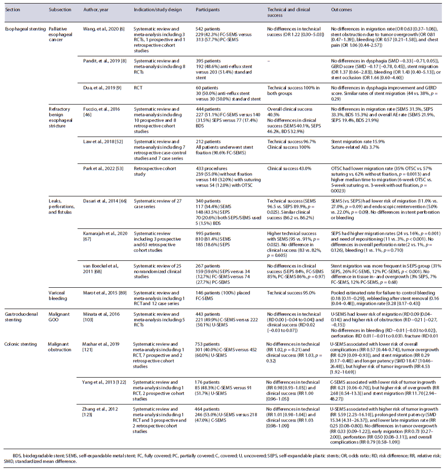
A meta-analysis of 18 studies did not show significant differences in clinical success, stent migration, and complication rates between BDS, SEMS, or SEPS [46]. However, patients with BDS (Fig. 2) may require fewer endoscopic reinterventions [56-58]. Despite this, updated European Society of Gastrointestinal Endoscopy (ESGE) guidelines do not recommend BDS over other stents [26]. Lumen-apposing metal stents (LAMSs) also have been evaluated for RBES, but available data are limited to small case series [59-62]. They may be considered in patients with short RBES up to 10 mm (Fig. 3). In patients with persistent dysphagia despite stent placement, surgery should be considered. Self-dilatation with boogies may be an option for poor surgical candidates [63].

Recommendation: Temporary placement of self-expandable stents may be considered for RBES. No recommendation can be made regarding a specific type of ex-pandable stent. When SEMSs are used, FC-SEMS should be preferred. Stent fixation techniques can be used to mitigate migration risk.

Fig. 2 Patient with a refractory benign esophageal stricture due to caustic ingestion, submitted to multiple endoscopic treatments (Savary and balloon dilatation, fully covered self-expandable metal stent placement). a Endoscopic image of esophageal stricture. b Fluoroscopic image revealing a 2-cm-long stricture after contrast instillation. c-f Endoscopic and fluoroscopic images after placement of a 25/20/25 × 100-mm biodegradable noncovered stent. 

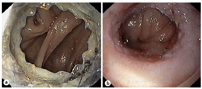
Fig. 3 Patient with a refractory esophago-jejunal anastomotic stricture who under-went placement of a lumen-apposing metal stent (LAMS) across the stricture. a Endoscopic image of the LAMS placed across the stricture. b Esophagojejunal anastomotic stricture remodeling after LAMS removal. 

Leaks, Perforations, and Fistulas

Recent advances in endoscopy have prompted a paradigm shift in the management of esophageal leaks, perforations, and fistulas, from surgery to minimally invasive endoscopic approaches [64]. Even though these terms are often used interchangeably, in strict terms, they are completely different [65]. Therefore, their treatment should be individualized.

Based on three systematic reviews on the use of PC-SEMS, FC-SEMS, and SEPS in anastomotic leaks and perforations, the clinical success rate of esophageal stent placement is 81-87%, with no difference among the stent types [66-68]. Only two studies [69, 70] evaluated fistulas individually, with clinical success ranging from 45.5 to 90.1%; however, SEMSs were used almost always in combination with other endoscopic/pulmonary techniques; clinical success decreased with orifice size increase [69]. Huh et al. [71] and Suzuki et al. [72] reported higher clinical success for perforations compared to leaks (100% vs. 60-80%), with anastomotic leak group needing a longer stent dwell time (≥4 weeks) compared with the perforation group (75% vs. 27.3%, p = 0.022). Overall AEs ranged from 3.8 to 50% [70, 71, 73, 74], with stent migration (8.5-42%) [67, 74-78] and strictures or stentinduced ulcers (3-48%) [73, 79] being the commonest. Even though stent-related AEs are typically managed endoscopically, severe AEs (14.7%) [80] can occur, requiring nonendoscopic advanced management.

The selection of the right stent design also remains a challenge (Fig. 4). Even though clinical success rates are comparable, SEMSs perform better than SEPS in leaks and perforations, with higher technical success (95% vs. 91%, p = 0.032), reduced risk of migration (16% vs. 24%, p = 0.001), and need for stent repositioning (3% vs. 11%, p < 0.001), as well as lower risk of perforation when considering anastomotic leaks only (0% vs. 2%, p = 0.013) [67]. Migration rates are higher with FC-SEMS versus PC-SEMS (odds ratio [OR] 2.44, 95% CI 1.13-5.31; p = 0.024) [77]; however, suturing FC-SEMS may render migration rates similar to PC-SEMS (adjusted OR 0.56, 95% CI 0.15-2.00; p = 0.37), without the difficulties in removal of PC-SEMS and a lower risk of AEs (21% vs. 46%, p = 0.37) [51]. Shim technique (silk thread attached to proximal end of the stent and to the patient ear via the nares) [81] as well as stents with wider diameters [77, 82] may also result in lower migration rates. Data regarding the role of BDS in management of esophageal transmural defects are limited. Only two studies, comprising 13 and 4 patients, are available: despite a clinical success of 77.8-100%, mucosal reaction (2/4 patients) is a drawback, causing dysphagia and requiring endoscopic dilation [83, 84].

Fig. 4 Patient with an anastomotic leak after total gastrectomy. a Endoscopic image showing an anastomotic leak occupying more than 50% of the luminal circumference. b Immediately after placement of a fully covered self-expandable metal stent. 

Predictive factors for stent failure/mortality include persistence of fistula orifice after 6 months of endoscopic treatment (OR 44, 95% CI 3.38-573.4; p = 0.004) [69], larger fistula size [69], if stent was used after failure of revisional therapy compared with stent used as initial treatment (55% vs. 100%, p = 0.013) [73], continuous leakage after stent placement [85], decreased physical performance preoperatively [85], and concomitant esophagotracheal fistula [85]. Van Halsema et al. [86] developed a prediction rule for successful stent placement in the context of benign upper GI leakage, consisting of etiology, location, size of the leak, and C-reactive protein level at diagnosis. Iatrogenic/spontaneous perforation (vs. leaks or fistulas), proximal defect location (<25 cm from the incisive), lower C-reactive protein levels, and smaller defect sizes (<1 cm) were considered predictors of better outcomes; after validation in a different patient cohort, the rule was found to significantly discriminate between failure (NPV 86%) and success (PPV 87%) of stent placement in patients with a predicted low (≤50%) or high (≥70%) clinical success, respectively.

ESGE-updated guidelines recommend removing the stent 6-8 weeks after placement [26], even though there is a tendency to remove or replace stents at shorter interval times, to reduce stent-related AEs. SEMSs have been compared to endoscopic vacuum therapy for the treatment of post-surgical leaks in two systematic reviews and meta-analyses [87, 88], with endoscopic vacuum therapy being associated with higher leak closure, more endoscopic device changes, shorter duration of treatment, and lower rates of mortality and/or major complications. Given the high complexity and particularities of transmural defects, in most cases a multimodality approach is adopted, but endoscopic stenting remains one of the most frequently used options in these patients [77].

Recommendation: Temporary SEMS placement can be considered for leaks, perforations, and fistulae. Considering the complexity of these transmural defects, a multi-modality approach is often preferred.

Acute Variceal Bleeding

In the setting of refractory acute variceal bleeding, several systematic reviews and meta-analyses [89-91] support use of SEMS in successful control of severe or refractory acute variceal bleeding, without significant device-related AEs (Fig. 5). This strategy is often used as a bridge to transjugular intrahepatic portosystemic shunt or liver transplantation in a significant proportion of patients [89], and 6-week survival is mostly related to the severity of the underlying liver disease. Dedicated FC-SEMSs (SX-Ella Danis) for esophageal variceal bleeding are available; when used, retrieval should be performed using a specifically designed system [92, 93]. There is only one RCT comparing FC-SEMS (SX-Ella Danis stent) with balloon tamponade [94], with successful therapy more frequent in the stent group (66% vs. 20%), with a significantly higher rate for control of bleeding (85% vs. 47%), lower transfusion requirements, and a lower incidence of seri-ous AEs (15% vs. 47%), mainly due to differences in aspiration pneumonia (0 vs. 5) and esophageal tear (1 patient in the balloon tamponade group); no significant difference in 6-week survival was observed (54% vs. 40%). In most published studies, FC-SEMSs were left in place for up to 2 weeks [95-98], although extended dwell time up to 30 days has been reported. Recommendation: FC-SEMS placement may be considered for the treatment of severe or refractory esophageal variceal bleeding, as a bridge to transjugular intrahepatic portosystemic shunt or liver transplantation.

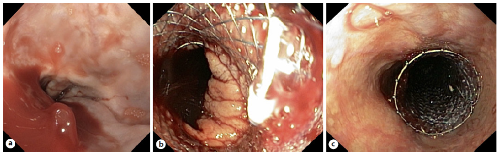
Fig. 5 Patient with cirrhosis Child-Pugh C and severe esophageal variceal bleeding. a Endoscopic image showing active bleeding from an esophageal varix. b, c Endoscopic image after placement of a dedicated fully covered self-expandable metal stent (SX-Ella Danis stent) with bleeding control. 

Gastroduodenal Stenting

GOO typically involves the distal stomach and/or the proximal small bowel (although it may also affect the distal small bowel) and may be secondary to mechanical/obstructive or motility causes. Mechanical obstructions can be benign or malignant [99]. The traditional approach for management of malignant GOO involves surgical gastrojejunostomy, via either open or laparoscopic access, although less invasive alternatives including endoscopic placement of luminal SEMS (Fig. 6) and, more recently, endoscopic ultrasound (EUS)-guided gastroenterostomies have become increasingly popular. On the contrary, benign GOO is generally managed with endoscopic balloon dilation (EBD), reserving more invasive techniques for EBD refractory cases [100].

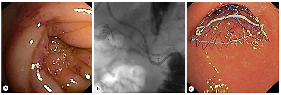
Fig. 6 a Patient with pancreatic cancer and previous biliary self-expandable metal stent with gastric outlet ob-struction due to tumor invasion in the second portion of the duodenum. b, c Fluoroscopic and endoscopic images after placement of an uncovered 140 × 20 mm self-expandable metal stent. 

A recent meta-analysis favored surgical gastrojejunostomy over SEMS due to longer overall survival and fewer needs for reintervention. Although postoperative mortality and AEs were similar between the two groups, the SEMS group had shorter hospital stay and shorter time to resume oral intake [101]. Technical and clinical success was 83.3-100% and 75-100%, respectively [101]. Therefore, patients with short life expectancy (<6 months), especially those who are high surgical risk, may be better candidates for luminal SEMS [102]. Regarding the stent type for GOO, a 2016 meta-analysis noted no significant difference in technical or clinical success, AEs, and reintervention for covered SEMS (C-SEMS) and uncovered SEMS (U-SEMS), but as expected, migration rate was higher, while obstruction rate was lower with C-SEMS [103].

In patients with combined malignant duodenal and biliary obstruction, “double stenting” should be the standard of care practice, due to its lower invasiveness and shorter recovery time [104]. Regarding approach for biliary stenting, endoscopic retrograde cholangiopancreatography stenting might be associated with a lower AE rate compared to EUS-guided biliary drainage and should be considered the preferred approach, when feasible [105].

Only a few case series have been published regarding SEMS as salvage therapy for benign GOO who failed initial EBD attempt. Despite symptomatic improvement in almost 80% of the patients, SEMS placement is limited by stent migration rates up to 47% [106, 107], with no robust evidence to recommend SEMS over surgery in these patients [102].

Recommendation: In patients with life expectancy below 6 months, especially if at high surgical risk, luminal SEMS can be considered. Otherwise, gastroenteric anastomosis should be considered, either surgical or endoscopic. Combined malignant duodenal and biliary obstruction should be approached with “double stenting.”

Colonic Stenting

Malignant Colonic Obstruction

Colonic stenting is a valid alternative to emergency surgery in patients with malignant colonic obstruction, either as bridge to surgery or palliative intention. Prophylactic stenting, in the absence of symptomatic obstruction, should not be performed. Most of the literature concerns left-sided obstructing colon cancer (Fig. 7), excluding (distal) rectal cancers; however, SEMS may also be successfully placed in malignant obstruction of the proximal/right colon [108]. As a bridge to surgery, colonic stenting is associated with fewer overall AEs, similar 30-day mortality rate, and a higher proportion of primary anastomoses, compared to emergency surgery. Even though pre-surgical colon stenting (as bridge therapy) may be associated with a higher overall tumor recurrence, this did not translate into a significant difference in terms of disease-free survival or overall survival on 3- and 5-year follow-up [109, 110]. The worse oncologic outcomes seem to be explained by stent-related perforations, with overall survival being better in studies with lower perforation rates. The ideal time interval for surgery after colonic stenting should be balanced between stent-related AEs (reduced by a short interval) and surgical outcomes (improved by a longer interval). ESGE-updated guideline suggests a 2-week interval between stent placement and surgery [111]. In patients who are not good candidates for colonic stenting (locally advanced disease requiring neo-adjuvant therapy or longer stenosis) or who fail stent placement, a decompressing stoma may be an alternative as a bridge to surgery, allowing a higher chance of successful primary anastomosis [112].

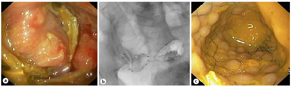
Fig. 7 Patient with malignant colonic obstruction due to colorectal cancer. a, b Fluoroscopic and endoscopic images after placement of an uncovered 80 × 20 mm self-expandable metal stent. 

In a palliative setting, most meta-analyses have demonstrated SEMS to be associated with lower short-term mortality, hospital stay, early AEs, stoma rates, and time to initiation of chemotherapy, compared to emergency surgery. Conversely, late AEs were more frequent in the SEMS group [113-117]. Chemotherapy does not seem to be a risk factor for colonic stent-related complications in general; however, in patients already receiving bevacizumab, stent placement is not advised due to high risk of perforation [111].

Extra-colonic malignancy complicated with colonic obstruction may benefit from palliative colonic stenting and is associated with fewer AEs compared to decompressive surgery. Unfortunately, technical and clinical success rates are lower compared to primary colonic cancer [118-120].

In terms of type of colonic stents, 3 meta-analyses have compared U-SEMS and C-SEMS, noting similar technical and clinical success, but U-SEMSs were associated with fewer overall AEs, including less tumor overgrowth, lower migration rates, longer patency, and fewer reinsertions, although at the cost of higher risk of tumor in-growth [121-123]. The main complications included perforation, stent failure, stent migration, and stent re-obstruction [124]. Migration should be treated with stent replacement or SIS technique in the palliative setting, and early surgery in the bridge-to-surgery patients [125].

Recommendation: SEMS can be considered for malignant colonic obstruction treatment as bridge to surgery (advantages and disadvantages of its placement must be discussed with the patient) or in palliative setting. Despite lower success rate, SEMS can also be used for extra-colonic malignant obstruction treatment.

Benign Colonic Obstruction

In recent years, the use of SEMS has been extended to treatment of benign GI strictures secondary to diverticulitis, radiation colitis, inflammatory bowel disease, and endometriosis, as well to management of post-anastomotic colonic leaks, strictures, and fistulas [126]. However, majority of data available in this regard are derived from retrospective studies.

Several studies have reported outcomes of colonic stenting for diverticulitis-associated strictures, as a bridge to surgery or for palliation (in poor surgical candidates). A systematic review (n = 66) concluded that the AE rate was not acceptable to warrant its use (11/66 perforations) [127]. Regarding fibrostenosic Crohn’s disease (CD) refractory to medical treatment, SEMS use is only described in small case series [128]. The largest case series, with a stent dwell time of 4 weeks, showed treatment efficacy of 64.7%, with one AE (proximal stent migration). Distal stent migration (52%) was not considered an AE but rather an incident [129]. A systematic review evaluated SEMS placement for the management of colorectal surgical complications including anastomotic strictures, leaks, or fistulas. A high early success rate (73.3%) was observed; however, anastomotic strictures were more challenging to treat, as around 50% of the patients had persistent stenosis and 26% required EBD after stent placement [130]. Complications were reported in 41.5% patients, mainly SEMS migration, explained by the inherent characteristics of C-SEMS [131]. Colonic stent placement in bowel obstruction due to endo-metriosis, colonic fistulas, radiation-induced stenosis, or ischemic colitis is also reported in literature, but only as case reports or short case series [126, 127].

The largest case series of BDS in colon and ileocolic anastomotic strictures report a technical success of 90-100% but only a modest stricture resolution of 45-83%. Unlike in esophageal strictures, mucosal hyperplastic reaction after BDS placement has not been reported in intestinal strictures [132, 133]. Use of BDS for CD strictures can theoretically overcome the shortcomings of SEMS (stent migration and need for stent removal); however, absence of biodegradable through-the-scope colonic stents makes deployment proximal to the sigmoid technically challenging. Data are very limited in this context. A case series of 11 BDS for treatment of CD strictures of the terminal ileum or colon (deployed through overtube, assisted by a stiff guidewire, and fluoroscopy guidance) revealed high technical success (90.9%), but early stent migration occurred in 3 patients [134].

Henceforth, limited available data do not support endorsement of SEMS placement in the context of benign colonic conditions and should only be considered in case-by-case basis after multidisciplinary discussion. Recom-mendation: SEMS placement in benign colonic strictures should not be routinely performed.

Conclusion

Endoscopic stenting practices and techniques are continuously evolving, requiring clinicians to be aware of updated evidence in this field (Table 2). For patients with unresectable esophageal cancer, SEMS placement is recommended as a palliative measure if expected survival is less than or equal to 3 months. If available, brachytherapy should be considered as an adjunct for patients with expected survival above 3 months. SEMS placement is also recommended for patients with malignant tracheoesophageal fistulas as well as patients with RBES and transmural defects. Gastroduodenal stenting should be considered in patients with malignant GOO, especially those who have a short life expectancy (below 6 months). Colonic SEMS is the preferred treatment for palliation of malignant colonic obstruction and can be considered as bridge to surgery in selected patients. In all cases, individualized considerations and the multidisciplinary context should be made when developing management recommendations and plans.

Table 2 Summary of main indications for esophageal, gastroduodenal, and colonic stenting and recommended types of stents for each indication 

References

1. Kalloo AN. Evolution of surgery to endoscopy. Curr Opin Gastroenterol. 2018; 34(5): 281. [ Links ]

2. ASGE Technology Committee; Varadarajulu S, Banerjee S, Barth B, Wang A, Desilets D, et al. Enteral stents. Gastrointest Endosc. 2011; 74(3): 455-64. [ Links ]

3. McLoughlin MT, Byrne MF. Endoscopic stenting: where are we now and where can we go? World J Gastroenterol. 2008; 14(24): 3798-803. [ Links ]

4. Perumpail RB, Muthusamy VR. Gastrointestinal stents: thinking outside the box. Tech Innov Gastrointest Endosc. 2020; 22(4): 239-44. [ Links ]

5. Sreedharan A, Harris K, Crellin A, Forman D, Everett SM. Interventions for dysphagia in oesophageal cancer. Cochrane Database Syst Rev. 2009; 2014(10): Cd005048. [ Links ]

6. Wang C, Wei H, Li Y. Comparison of fullycovered vs. partially covered self-expanding metallic stents for palliative treatment of inoperable esophageal malignancy: a systematic review and meta-analysis. BMC Cancer. 2020; 20(1): 73. [ Links ]

7. Spaander MCW, Baron TH, Siersema PD, Fuccio L, Schumacher B, Escorsell À, et al. Esophageal stenting for benign and malignant disease: European Society of Gastrointestinal Endoscopy (ESGE) Clinical Guideline. Endoscopy. 2016; 48(10): 939-48. [ Links ]

8. Pandit S, Samant H, Morris J, Alexander SJ. Efficacy and safety of standard and anti-reflux self-expanding metal stent: a systematic review and meta-analysis of randomized controlled trials. World J Gastrointest Endosc. 2019; 11(4): 271-80. [ Links ]

9. Dua KS, DeWitt JM, Kessler WR, Diehl DL, Draganov PV, Wagh MS, et al. A phase III, multicenter, prospective, single-blinded, noninferiority, randomized controlled trial on the performance of a novel esophageal stent with an antireflux valve (with video). Gastrointest Endosc. 2019; 90(1): 64-74.e3. [ Links ]

10. Homs MYV, Steyerberg EW, Eijkenboom WMH, Tilanus HW, Stalpers LJA, Bartelsman JFWM, et al. Single-dose brachytherapy versus metal stent placement for the palliation of dysphagia from oesophageal cancer: multicentre randomised trial. Lancet. 2004; 364(9444): 1497-504. [ Links ]

11. Bergquist H, Wenger U, Johnsson E, Nyman J, Ejnell H, Hammerlid E, et al. Stent insertion or endoluminal brachytherapy as palliation of patients with advanced cancer of the esophagus and gastroesophageal junction. Results of a randomized, controlled clinical trial. Dis Esophagus. 2005; 18(3): 131-9. [ Links ]

12. Fuccio L, Guido A, Hassan C, Frazzoni L, Arcelli A, Farioli A, et al. Underuse of brachytherapy for the treatment of dysphagia owing to esophageal cancer. An Italian survey. Dig Liver Dis. 2016; 48(10): 1233-6. [ Links ]

13. Salembier C, De Hertogh O, Daisne JF, Palumbo S, Van Gestel D. Brachytherapy in Belgium in 2018. A national survey of the brachytherapy study group of the Belgian SocieTy for Radiotherapy and Oncology (BeSTRO). Radiother Oncol. 2020; 150: 245-52. [ Links ]

14. Fuccio L, Mandolesi D, Farioli A, Hassan C, Frazzoni L, Guido A, et al. Brachytherapy for the palliation of dysphagia owing to esophageal cancer: a systematic review and metaanalysis of prospective studies. Radiother Oncol. 2017; 122(3): 332-9. [ Links ]

15. Lai A, Lipka S, Kumar A, Sethi S, Bromberg D, Li N, et al. Role of esophageal metal stents placement and combination therapy in inoperable esophageal carcinoma: a systematic review and meta-analysis. Dig Dis Sci. 2018; 63(4): 1025-34. [ Links ]

16. Wong AT, Shao M, Rineer J, Osborn V, Schwartz D, Schreiber D. Treatment and survival outcomes of small cell carcinoma of the esophagus: an analysis of the National Cancer Data Base. Dis Esophagus. 2017; 30(2): 1-5. [ Links ]

17. Martin EJ, Bruggeman AR, Nalawade VV, Sarkar RR, Qiao EM, Rose BS, et al. Palliative radiotherapy versus esophageal stent placement in the management of patients with metastatic esophageal cancer. J Natl Compr Canc Netw. 2020; 18(5): 569-74. [ Links ]

18. Jeene PM, Vermeulen BD, Rozema T, Braam PM, Lips I, Muller K, et al. Short-course external beam radiotherapy versus brachytherapy for palliation of dysphagia in esophageal cancer: a matched comparison of two prospective trials. J Thorac Oncol. 2020; 15(8): 1361-8. [ Links ]

19. Penniment MG, De Ieso PB, Harvey JA, Stephens S, Au HJ, O'Callaghan CJ, et al. Palliative chemoradiotherapy versus radiotherapy alone for dysphagia in advanced oesophageal cancer: a multicentre randomised controlled trial (TROG 03.01). Lancet Gastroenterol Hepatol. 2018; 3(2): 114-24. [ Links ]

20. Bakheet N, Park JH, Hu HT, Yoon SH, Kim KY, Zhe W, et al. Fully covered self-expandable esophageal metallic stents in patients with inoperable malignant disease who survived for more than 6 months after stent Placement. Br J Radiol. 2019; 92(1100): 20190321. [ Links ]

21. Medeiros VS, Martins BC, Lenz L, Ribeiro MSI, de Paulo GA, Lima MS, et al. Adverse events of self-expandable esophageal metallic stents in patients with long-term survival from advanced malignant disease. Gastrointest Endosc. 2017; 86(2): 299-306. [ Links ]

22. Rodrigues-Pinto E, Pereira P, Baron TH, Macedo G. Self-expandable metal stents are a valid option in long-term survivors of advanced esophageal cancer. Rev Esp Enferm Dig. 2018; 110(8): 500-4. [ Links ]

23. Chen HL, Shen WQ, Liu K. Radioactive selfexpanding stents for palliative management of unresectable esophageal cancer: a systematic review and meta-analysis. Dis Esophagus. 2017; 30(5): 1-16. [ Links ]

24. Yang ZM, Geng HT, Wu H. Radioactive stent for malignant esophageal obstruction: a meta-analysis of randomized controlled trials. J Laparoendosc Adv Surg Tech A. 2021; 31(7): 783-9. [ Links ]

25. Ferreira-Silva J, Medas R, Girotra M, Barakat M, Tabibian JH, Rodrigues-Pinto E. Futuristic developments and applications in endoluminal stenting. Gastroenterol Res Pract. 2022; 2022: 6774925. [ Links ]

26. Spaander MCW, van der Bogt RD, Baron TH, Albers D, Blero D, de Ceglie A, et al. Esophageal stenting for benign and malignant disease: European Society of Gastrointestinal Endoscopy (ESGE) Guideline - update 2021. Endoscopy. 2021; 53(7): 751-62. [ Links ]

27. Ahmed O, Bolger JC, O'Neill B, Robb WB. Use of esophageal stents to relieve dysphagia during neoadjuvant therapy prior to esophageal resection: a systematic review. Dis Esophagus. 2020; 33(1): doz090. [ Links ]

28. Helminen O, Kauppila JH, Kytö V, Gunn J, Lagergren J, Sihvo E. Preoperative esophageal stenting and short-term outcomes of surgery for esophageal cancer in a population-based study from Finland and Sweden. Dis Esophagus. 2019; 32(11): doz005. [ Links ]

29. Järvinen T, Ilonen I, Ylikoski E, Nelskylä K, Kauppi J, Salo J, et al. Preoperative stenting in oesophageal cancer has no effect on survival: a propensity-matched case-control study. Eur J Cardiothorac Surg. 2017; 52(2): 385-91. [ Links ]

30. Rodrigues-Pinto E, Ferreira-Silva J, Sousa-Pinto B, Medas R, Garrido I, Siersema PD, et al. Self-expandable metal stents in esophageal cancer before preoperative neoadjuvant therapy: efficacy, safety, and long-term outcomes. Surg Endosc. 2021; 35(9): 5130-9. [ Links ]

31. Nagaraja V, Cox MR, Eslick GD. Safety and efficacy of esophageal stents preceding or during neoadjuvant chemotherapy for esophageal cancer: a systematic review and metaanalysis. J Gastrointest Oncol. 2014; 5(2): 119-26. [ Links ]

32. Bower M, Jones W, Vessels B, Scoggins C, Martin R. Nutritional support with endoluminal stenting during neoadjuvant therapy for esophageal malignancy. Ann Surg Oncol. 2009; 16(11): 3161-8. [ Links ]

33. Siddiqui AA, Glynn C, Loren D, Kowalski T. Self-expanding plastic esophageal stents versus jejunostomy tubes for the maintenance of nutrition during neoadjuvant chemoradiation therapy in patients with esophageal cancer: a retrospective study. Dis Esophagus. 2009; 22(3): 216-22. [ Links ]

34. Mariette C, Gronnier C, Duhamel A, Mabrut JY, Bail JP, Carrere N, et al. Self-expanding covered metallic stent as a bridge to surgery in esophageal cancer: impact on oncologic outcomes. J Am Coll Surg. 2015; 220(3): 287-96. [ Links ]

35. Martin R, Duvall R, Ellis S, Scoggins CR. The use of self-expanding silicone stents in esophageal cancer care: optimal pre-peri-and postoperative care. Surg Endosc. 2009; 23(3): 615-21. [ Links ]

36. Shamji FM, Inculet R. Management of malignant tracheoesophageal fistula. Thorac Surg Clin. 2018; 28(3): 393-402. [ Links ]

37. Aworanti O, Awadalla S. Management of recurrent tracheoesophageal fistulas: a systematic review. Eur J Pediatr Surg. 2014; 24(5): 365-75. [ Links ]

38. Ramai D, Bivona A, Latson W, Ofosu A, Ofori E, Reddy M, et al. Endoscopic management of tracheoesophageal fistulas. Ann Gastroenterol. 2019; 32(1): 24-9. [ Links ]

39. Ke M, Wu X, Zeng J. The treatment strategy for tracheoesophageal fistula. J Thorac Dis. 2015; 7(Suppl 4): S389-97. [ Links ]

40. Laasch HU, Najran P, Mullan D, Shepherd D, Li A, Najran PLaasch HU. Minimally invasive treatment strategies for tracheoesophageal fistulae. Dig Dis Interv. 2018; 2(1): 11-7. [ Links ]

41. Nomori H, Horio H, Imazu Y, Suemasu K. Double stenting for esophageal and tracheobronchial stenoses. Ann Thorac Surg. 2000; 70(6): 1803-7. [ Links ]

42. Binkert CA, Petersen BD. Two fatal complications after parallel tracheal-esophageal stenting. Cardiovasc Intervent Radiol. 2002; 25(2): 144-7. [ Links ]

43. Hürtgen M, Herber SCA. Treatment of malignant tracheoesophageal fistula. Thorac Surg Clin. 2014; 24(1): 117-27. [ Links ]

44. Reed MF, Mathisen DJ. Tracheoesophageal fistula. Chest Surg Clin N Am. 2003; 13(2): 271-89. [ Links ]

45. Herth FJF, Peter S, Baty F, Eberhardt R, Leuppi JD, Chhajed PN. Combined airway and oesophageal stenting in malignant airway-oesophageal fistulas: a prospective study. Eur Respir J. 2010; 36(6): 1370-4. [ Links ]

46. Fuccio L, Hassan C, Frazzoni L, Miglio R, Repici A. Clinical outcomes following stent placement in refractory benign esophageal stricture: a systematic review and meta-analysis. Endoscopy. 2016; 48(2): 141-8. [ Links ]

47. Wang C, Lou C. Randomized controlled trial to investigate the effect of metal clips on early migration during stent implantation for malignant esophageal stricture. Can J Surg. 2015; 58(6): 378-82. [ Links ]

48. Conio M, Savarese MF, Baron TH, De Ceglie A. A newly designed over-the-scope-clip device to prevent fully covered metal stents migration: a pilot study. Tech Innov Gastrointest Endosc. 2020; 22(4): 167-71. [ Links ]

49. Bick BL, Imperiale TF, Johnson CS, DeWitt JM. Endoscopic suturing of esophageal fully covered self-expanding metal stents reduces rates of stent migration. Gastrointest Endosc. 2017; 86(6): 1015-21. [ Links ]

50. Sharaiha RZ, Kumta NA, Doukides TP, Eguia V, Gonda TA, Widmer JL, et al. Esophageal stenting with sutures: time to redefine our standards? J Clin Gastroenterol. 2015; 49(6): e57-60. [ Links ]

51. Ngamruengphong S, Sharaiha R, Sethi A, Siddiqui A, DiMaio CJ, Gonzalez S, et al. Fullycovered metal stents with endoscopic suturing versus. partially-covered metal stents for benign upper gastrointestinal diseases: a comparative study. Endosc Int Open. 2018; 6(2): E217-23. [ Links ]

52. Law R, Prabhu A, Fujii-Lau L, Shannon C, Singh S. Stent migration following endoscopic suture fixation of esophageal self-expandable metal stents: a systematic review and meta- analysis. Surg Endosc. 2018; 32(2): 675-81. [ Links ]

53. Park KH, Lew D, Samaan J, Patel S, Liu Q, Gaddam S, et al. Comparison of no stent fixation, endoscopic suturing, and a novel overthe-scope clip for stent fixation in preventing migration of fully covered self expanding metal stents: a retrospective comparative study (with video). Gastrointest Endosc. 2022. Epub ahead of print. [ Links ]

54. van Halsema EE, Wong Kee Song LM, Baron TH, Siersema PD, Vleggaar FP, Ginsberg GG, et al. Safety of endoscopic removal of self-expandable stents after treatment of benign esophageal diseases. Gastrointest Endosc. 2013; 77(1): 18-28. [ Links ]

55. DaVee T, Irani S, Leggett CL, Berzosa Corella M, Grooteman KV, Wong Kee Song LM, et al. Stent-in-stent technique for removal of embedded partially covered self-expanding metal stents. Surg Endosc. 2016; 30(6): 2332-41. [ Links ]

56. van Boeckel PGA, Vleggaar FP, Siersema PD. A comparison of temporary self-expanding plastic and biodegradable stents for refractory benign esophageal strictures. Clin Gastroenterol Hepatol. 2011; 9(8): 653-9. [ Links ]

57. Canena JMT, Liberato MJA, Rio-Tinto RAN, Pinto-Marques PM, Romão CMM, Coutinho AVMP, et al. A comparison of the temporary placement of 3 different self-expanding stents for the treatment of refractory benign esophageal strictures: a prospective multicentre study. BMC Gastroenterol. 2012; 12: 70. [ Links ]

58. Walter D, van den Berg MW, Hirdes MM, Vleggaar FP, Repici A, Deprez PH, et al. Dilation or biodegradable stent placement for recurrent benign esophageal strictures: a randomized controlled trial. Endoscopy. 2018; 50(12): 1146-55. [ Links ]

59. Adler DG. Lumen-apposing metal stents for the treatment of refractory Benign esophageal strictures. Am J Gastroenterol. 2017; 112(3): 516-7. [ Links ]

60. Irani S, Jalaj S, Ross A, Larsen M, Grimm IS, Baron TH. Use of a lumen-apposing metal stent to treat GI strictures (with videos). Gastrointest Endosc. 2017; 85(6): 1285-9. [ Links ]

61. Santos-Fernandez J, Paiji C, Shakhatreh M, Becerro-Gonzalez I, Sanchez-Ocana R, Yeaton P, et al. Lumen-apposing metal stents for benign gastrointestinal tract strictures: an international multicenter experience. World J Gastrointest Endosc. 2017; 9(12): 571-8. [ Links ]

62. Yang D, Nieto JM, Siddiqui A, Riff BP, Di-Maio CJ, Nagula S, et al. Lumen-apposing covered self-expandable metal stents for short benign gastrointestinal strictures: a multicenter study. Endoscopy. 2017; 49(4): 327-33. [ Links ]

63. van Halsema EE, t Hoen CA, de Koning PS, Rosmolen WD, van Hooft JE, Bergman JJ. Self-dilation for therapy-resistant benign esophageal strictures: towards a systematic approach. Surg Endosc. 2018; 32(7): 3200-7. [ Links ]

64. Cereatti F, Grassia R, Drago A, Conti CB, Donatelli G. Endoscopic management of gastrointestinal leaks and fistulae: what option do we have? World J Gastroenterol. 2020; 26(29): 4198-217. [ Links ]

65. Fernández-Urién I, Vila J. Esophageal leaks: i thought that glue was not effective. Endosc Int Open. 2018; 6(9): E1100-2. [ Links ]

66. Dasari BVM, Neely D, Kennedy A, Spence G, Rice P, Mackle E, et al. The role of esophageal stents in the management of esophageal anastomotic leaks and benign esophageal perforations. Ann Surg. 2014; 259(5): 852-60. [ Links ]

67. Kamarajah SK, Bundred J, Spence G, Kennedy A, Dasari BVM, Griffiths EA. Critical appraisal of the impact of oesophageal stents in the management of oesophageal anastomotic leaks and Benign oesophageal perforations: an updated systematic review. World J Surg. 2020; 44(4): 1173-89. [ Links ]

68. van Boeckel PGA, Sijbring A, Vleggaar FP, Siersema PD. Systematic review: temporary stent placement for benign rupture or anastomotic leak of the oesophagus. Aliment Pharmacol Ther. 2011; 33(12): 1292-301. [ Links ]

69. Debourdeau A, Gonzalez JM, Dutau H, Benezech A, Barthet M. Endoscopic treatment of nonmalignant tracheoesophageal and bronchoesophageal fistula: results and prognostic factors for its success. Surg Endosc. 2019; 33(2): 549-56. [ Links ]

70. Silon B, Siddiqui AA, Taylor LJ, Arastu S, Soomro A, Adler DG. Endoscopic management of esophagorespiratory fistulas: a multicenter retrospective study of techniques and outcomes. Dig Dis Sci. 2017; 62(2): 424-31. [ Links ]

71. Huh CW, Kim JS, Choi HH, Lee JI, Ji JS, Kim BW, et al. Treatment of benign perforations and leaks of the esophagus: factors associated with success after stent placement. Surg Endosc. 2018; 32(8): 3646-51. [ Links ]

72. Suzuki T, Siddiqui A, Taylor LJ, Cox K, Hasan RA, Laique SN, et al. Clinical outcomes, efficacy, and adverse events in patients undergoing esophageal stent placement for Benign indications: a large multicenter study. J Clin Gastroenterol. 2016; 50(5): 373-8. [ Links ]

73. Boerlage TCC, Houben GPM, Groenen MJM, van der Linde K, van de Laar AWJM, Emous M, et al. A novel fully covered double-bump stent for staple line leaks after bariatric surgery: a retrospective analysis. Surg Endosc. 2018; 32(7): 3174-80. [ Links ]

74. van den Berg MW, Kerbert AC, van Soest EJ, Schwartz MP, Bakker CM, Gilissen LPL, et al. Safety and efficacy of a fully covered large-diameter self-expanding metal stent for the treatment of upper gastrointestinal perforations, anastomotic leaks, and fistula. Dis Esophagus. 2016; 29(6): 572-9. [ Links ]

75. Berlth F, Bludau M, Plum PS, Herbold T, Christ H, Alakus H, et al. Self-expanding metal stents versus endoscopic vacuum therapy in anastomotic leak treatment after oncologic gastroesophageal surgery. J Gastrointest Surg. 2019; 23(1): 67-75. [ Links ]

76. Okazaki O, Bernardo WM, Brunaldi VO, Junior CCd C, Minata MK, de Moura DTH, et al. Efficacy and safety of stents in the treatment of fistula after bariatric surgery: a systematic review and meta-analysis. Obes Surg. 2018; 28(6): 1788-96. [ Links ]

77. Rodrigues-Pinto E, Pereira P, Sousa-Pinto B, Shehab H, Pinho R, Larsen MC, et al. Retrospective multicenter study on endoscopic treatment of upper GI postsurgical leaks. Gastrointest Endosc. 2021; 93(6): 1283-99.e2. [ Links ]

78. Shehab H, Abdallah E, Gawdat K, Elattar I. Large bariatric-specific stents and over-thescope clips in the management of post-bariatric surgery leaks. Obes Surg. 2018; 28(1): 15-24. [ Links ]

79. Plum PS, Herbold T, Berlth F, Christ H, Alakus H, Bludau M, et al. Outcome of self-expanding metal stents in the treatment of anastomotic leaks after Ivor Lewis esophagectomy. World J Surg. 2019; 43(3): 862-9. [ Links ]

80. Bohle W, Louris I, Schaudt A, Koeninger J, Zoller WG. Predictors for treatment failure of self-expandable metal stents for anastomotic leak after gastro-esophageal resection. J Gastrointestin Liver Dis. 2020; 29(2): 145-9. [ Links ]

81. Choi CW, Kang DH, Kim HW, Park SB, Kim SJ, Hwang SH, et al. Full covered self-expandable metal stents for the treatment of anastomotic leak using a silk thread. Medicine. 2017; 96(29): e7439. [ Links ]

82. Sdralis EIK, Petousis S, Rashid F, Lorenzi B, Charalabopoulos A. Epidemiology, diagnosis, and management of esophageal perforations: systematic review. Dis Esophagus. 2017; 30(8): 1-6. [ Links ]

83. Al Lehibi A, Al Balkhi A, Al Mtawa A, Al Otaibi N. Endoscopic biodegradable stents as a rescue treatment in the management of post bariatric surgery leaks: a case series. Endosc Int Open. 2018; 6(6): E722-6. [ Links ]

84. Könes O, Oran E. Self-expanding biodegradable stents for postoperative upper gastrointestinal issues. JSLS. 2018; 22(2): e2018.00011. [ Links ]

85. Persson S, Rouvelas I, Kumagai K, Song H, Lindblad M, Lundell L, et al. Treatment of esophageal anastomotic leakage with self-expanding metal stents: analysis of risk factors for treatment failure. Endosc Int Open. 2016; 4(4): E420-6. [ Links ]

86. van Halsema EE, Kappelle WFW, Weusten BLAM, Lindeboom R, van Berge Henegouwen MI, Fockens P, et al. Stent placement for benign esophageal leaks, perforations, and fistulae: a clinical prediction rule for successful leakage control. Endoscopy. 2018; 50(2): 98-108. [ Links ]

87. Rausa E, Asti E, Aiolfi A, Bianco F, Bonitta G, Bonavina L. Comparison of endoscopic vacuum therapy versus endoscopic stenting for esophageal leaks: systematic review and metaanalysis. Dis Esophagus. 2018; 31(11). [ Links ]

88. Scognamiglio P, Reeh M, Karstens K, Bellon E, Kantowski M, Schön G, et al. Endoscopic vacuum therapy versus stenting for postoperative esophago-enteric anastomotic leakage: systematic review and meta-analysis. Endoscopy. 2020; 52(8): 632-42. [ Links ]

89. Marot A, Trépo E, Doerig C, Moreno C, Moradpour D, Deltenre P. Systematic review with meta-analysis: self-expanding metal stents in patients with cirrhosis and severe or refractory oesophageal variceal bleeding. Aliment Pharmacol Ther. 2015; 42(11-12):1250-60. [ Links ]

90. Mohan BP, Chandan S, Khan SR, Kotagiri R, Kassab LL, Olaiya B, et al. Self-expanding metal stents versus TIPS in treatment of refractory bleeding esophageal varices: a systematic review and meta-analysis. Endosc Int Open. 2020; 8(3): E291-300. [ Links ]

91. Rodrigues SG, Cárdenas A, Escorsell À, Bosch J. Balloon tamponade and esophageal stenting for esophageal variceal bleeding in cirrhosis: a systematic review and metaanalysis. Semin Liver Dis. 2019; 39(2): 178-94. [ Links ]

92. Pfisterer N, Riedl F, Pachofszky T, Gschwantler M, König K, Schuster B, et al. Outcomes after placement of a SX-ELLA oesophageal stent for refractory variceal bleeding: a national multicentre study. Liver Int. 2019; 39(2): 290-8. [ Links ]

93. Khan S, Gilhotra R, Di Jiang C, Rowbotham D, Chong A, Majumdar A, et al. The role of a novel self-expanding metal stent in variceal bleeding: a multicenter Australian and New Zealand experience. Endosc Int Open. 2022; 10(3): E238-45. [ Links ]

94. Escorsell À, Pavel O, Cárdenas A, Morillas R, Llop E, Villanueva C, et al. Esophageal balloon tamponade versus esophageal stent in controlling acute refractory variceal bleeding: a multicenter randomized, controlled trial. Hepatology. 2016; 63(6): 1957-67. [ Links ]

95. Escorsell A, Bosch J. Self-expandable metal stents in the treatment of acute esophageal variceal bleeding. Gastroenterol Res Pract. 2011; 2011: 910986. [ Links ]

96. Wright G, Lewis H, Hogan B, Burroughs A, Patch D, O'Beirne J. A self-expanding metal stent for complicated variceal hemorrhage: experience at a single center. Gastrointest Endosc. 2010; 71(1): 71-8. [ Links ]

97. Zehetner J, Shamiyeh A, Wayand W, Hubmann R. Results of a new method to stop acute bleeding from esophageal varices: implantation of a self-expanding stent. Surg Endosc. 2008; 22(10): 2149-52. [ Links ]

98. van Hooft JE, van Berge Henegouwen MI, Rauws EA, Bergman JJ, Busch OR, Fockens P. Endoscopic treatment of benign anastomotic esophagogastric strictures with a biodegradable stent. Gastrointest Endosc. 2011; 73(5): 1043-7. [ Links ]

99. Jeong SJ, Lee J. Management of gastric outlet obstruction: focusing on endoscopic approach. World J Gastrointest Pharmacol Ther. 2020; 11(2): 8-16. [ Links ]

100. Tringali A, Giannetti A, Adler DG. Endoscopic management of gastric outlet obstruction disease. Ann Gastroenterol. 2019; 32(4): 330-7. [ Links ]

101. Mintziras I, Miligkos M, Wächter S, Manoharan J, Bartsch DK. Palliative surgical bypass is superior to palliative endoscopic stenting in patients with malignant gastric outlet obstruction: systematic review and meta-analysis. Surg Endosc. 2019; 33(10): 3153-64. [ Links ]

102. ASGE Standards of Practice Committee; Jue TL, Storm AC, Naveed M, Fishman DS, Qumseya BJ, et al. ASGE guideline on the role of endoscopy in the management of benign and malignant gastroduodenal obstruction. Gastrointest Endosc. 2021; 93(2): 309-22.e4. [ Links ]

103. Minata MK, Bernardo WM, Rocha RS, Morita FHA, Aquino JCM, Cheng S, et al. Stents and surgical interventions in the palliation of gastric outlet obstruction: a systematic review. Endosc Int Open. 2016; 4(11): E1158-70. [ Links ]

104. Takeda T, Sasaki T, Okamoto T, Sasahira N. Endoscopic double stenting for the management of combined malignant biliary and duodenal obstruction. J Clin Med. 2021; 10(15): 3372. [ Links ]

105. Fábián A, Bor R, Gede N, Bacsur P, Pécsi D, Hegyi P, et al. Double stenting for malignant biliary and duodenal obstruction: a systematic review and meta-analysis. Clin Transl Gastroenterol. 2020; 11(4): e00161. [ Links ]

106. Choi WJ, Park JJ, Park J, Lim EH, Joo MK, Yun JW, et al. Effects of the temporary placement of a self-expandable metallic stent in benign pyloric stenosis. Gut Liver. 2013; 7(4): 417-22. [ Links ]

107. Heo J, Jung MK. Safety and efficacy of a partially covered self-expandable metal stent in benign pyloric obstruction. World J Gastroenterol. 2014; 20(44): 16721-5. [ Links ]

108. Amelung FJ, de Beaufort HWL, Siersema PD, Verheijen PM, Consten ECJ. Emergency resection versus bridge to surgery with stenting in patients with acute right-sided colonic obstruction: a systematic review focusing on mortality and morbidity rates. Int J Colorectal Dis. 2015; 30(9): 1147-55. [ Links ]

109. Foo CC, Poon SHT, Chiu RHY, Lam WY, Cheung LC, Law WL. Is bridge to surgery stenting a safe alternative to emergency surgery in malignant colonic obstruction: a meta-analysis of randomized control trials. Surg Endosc. 2019; 33(1): 293-302. [ Links ]

110. Spannenburg L, Sanchez Gonzalez M, Brooks A, Wei S, Li X, Liang X, et al. Surgical outcomes of colonic stents as a bridge to surgery versus emergency surgery for malignant colorectal obstruction: a systematic review and meta-analysis of high quality prospective and randomised controlled trials. Eur J Surg Oncol. 2020; 46(8): 1404-14. [ Links ]

111. van Hooft JE, Veld JV, Arnold D, Beets-Tan RGH, Everett S, Götz M, et al. Self-expandable metal stents for obstructing colonic and extracolonic cancer: European Society of Gastrointestinal Endoscopy (ESGE) Guideline - Update 2020. Endoscopy. 2020; 52(5): 389-407. [ Links ]

112. Veld JV, Amelung FJ, Borstlap WAA, van Halsema EE, Consten ECJ, Siersema PD, et al. Comparison of decompressing stoma versus stent as a bridge to surgery for leftsided obstructive colon cancer. JAMA Surg. 2020; 155(3): 206-15. [ Links ]

113. Veld J, Umans D, van Halsema E, Amelung F, Fernandes D, Lee MS, et al. Self-expandable metal stent (SEMS) placement or emergency surgery as palliative treatment for obstructive colorectal cancer: a systematic review and meta-analysis. Crit Rev Oncol Hematol. 2020; 155: 103110. [ Links ]

114. Liang TW, Sun Y, Wei YC, Yang DX. Palliative treatment of malignant colorectal obstruction caused by advanced malignancy: a self-expanding metallic stent or surgery? A system review and meta-analysis. Surg Today. 2014; 44(1): 22-33. [ Links ]

115. Ribeiro IB, Bernardo WM, Martins BDC, de Moura DTH, Baba ER, Josino IR, et al. Colonic stent versus emergency surgery as treatment of malignant colonic obstruction in the palliative setting: a systematic review and meta-analysis. Endosc Int Open. 2018; 6(5): E558-67. [ Links ]

116. Takahashi H, Okabayashi K, Tsuruta M, Hasegawa H, Yahagi M, Kitagawa Y. Selfexpanding metallic stents versus surgical intervention as palliative therapy for obstructive colorectal cancer: a meta-analysis. World J Surg. 2015; 39(8): 2037-44. [ Links ]

117. Zhao XD, Cai BB, Cao RS, Shi RH. Palliative treatment for incurable malignant colorectal obstructions: a meta-analysis. World J Gastroenterol. 2013; 19(33): 5565-74. [ Links ]

118. Ahn H, Yoon CJ, Lee JH, Choi WS. Stent placement for palliative treatment of malignant colorectal obstruction: extracolonic malignancy versus primary colorectal cancer. AJR Am J Roentgenol. 2020; 215(1): 248-53. [ Links ]

119. Kim JY, Kim SG, Im JP, Kim JS, Jung HC. Comparison of treatment outcomes of endoscopic stenting for colonic and extracolonic malignant obstruction. Surg Endosc. 2013; 27(1): 272-7. [ Links ]

120. Sano T, Nozawa Y, Iwanaga A, Azumi M, Imai M, Ishikawa T, et al. Comparison of the efficacy of self-expandable metallic stents in colorectal obstructions caused by extracolonic malignancy and colorectal cancer. Mol Clin Oncol. 2021; 15(2): 170. [ Links ]

121. Mashar M, Mashar R, Hajibandeh S. Uncovered versus covered stent in management of large bowel obstruction due to colorectal malignancy: a systematic review and metaanalysis. Int J Colorectal Dis. 2019; 34(5): 773-85. [ Links ]

122. Yang Z, Wu Q, Wang F, Ye X, Qi X, Fan D. A systematic review and meta-analysis of randomized trials and prospective studies comparing covered and bare self-expandable metal stents for the treatment of malignant obstruction in the digestive tract. Int J Med Sci. 2013; 10(7): 825-35. [ Links ]

123. Zhang Y, Shi J, Shi B, Song CY, Xie WF, Chen YX. Comparison of efficacy between uncovered and covered self-expanding metallic stents in malignant large bowel obstruction: a systematic review and metaanalysis. Colorectal Dis. 2012; 14(7): e367-74. [ Links ]

124. Frazzoni L, Fabbri E, Bazzoli F, Triantafyllou K, Fuccio L. Colorectal stenting for palliation and bridge to surgery of obstructing cancer. Tech Innov Gastrointest Endosc. 2020. [ Links ]

125. Yoon JY, Park SJ, Hong SP, Kim TI, Kim WH, Cheon JH. Outcomes of secondary self-expandable metal stents versus surgery after delayed initial palliative stent failure in malignant colorectal obstruction. Digestion. 2013; 88(1): 46-55. [ Links ]

126. Venezia L, Michielan A, Condino G, Sinagra E, Stasi E, Galeazzi M, et al. Feasibility and safety of self-expandable metal stent in nonmalignant disease of the lower gastrointestinal tract. World J Gastrointest Endosc. 2020; 12(2): 60-71. [ Links ]

127. Currie A, Christmas C, Aldean H, Mobasheri M, Bloom ITM. Systematic review of selfexpanding stents in the management of benign colorectal obstruction. Colorectal Dis. 2014; 16(4): 239-45. [ Links ]

128. Adamina M, Bonovas S, Raine T, Spinelli A, Warusavitarne J, Armuzzi A, et al. ECCO guidelines on therapeutics in Crohn's disease: surgical treatment. J Crohns Colitis. 2020; 14(2): 155-68. [ Links ]

129. Loras C, Pérez-Roldan F, Gornals JB, Barrio J, Igea F, González-Huix F, et al. Endoscopic treatment with self-expanding metal stents for Crohn's disease strictures. Aliment Pharmacol Ther. 2012; 36(9): 833-9. [ Links ]

130. Arezzo A, Bini R, Lo Secco G, Verra M, Passera R. The role of stents in the management of colorectal complications: a systematic review. Surg Endosc. 2017; 31(7): 2720-30. [ Links ]

131. Trovato C, Fiori G, Ravizza D, Tamayo D, Zampino MG, Biffi R, et al. Delayed colonic perforation after metal stent placement for malignant colorectal obstruction. Endoscopy. 2006; 38(Suppl 2): E96. [ Links ]

132. Pérez Roldán F, González Carro P, Villafáñez García MC, Aoufi Rabih S, Legaz Huidobro ML, Sánchez-Manjavacas Múñoz N, et al. Usefulness of biodegradable polydioxanone stents in the treatment of postsurgical colorectal strictures and fistulas. Endoscopy. 2012; 44(3): 297-300. [ Links ]

133. Janík V, Horák L, Hnanícek J, Málek J, Laasch HU. Biodegradable polydioxanone stents: a new option for therapy-resistant anastomotic strictures of the colon. Eur Radiol. 2011; 21(9): 1956-61. [ Links ]

134. Rejchrt S, Kopacova M, Brozik J, Bures J. Biodegradable stents for the treatment of benign stenoses of the small and large intestines. Endoscopy. 2011; 43(10): 911-7. [ Links ]

1Statement of Ethics The authors state that this article did not require ethics ap-proval, since it is a review article and does not involve animal or human studies.

3Funding Sources The authors have no funding sources to declare.

5Data Availability Statement Our study did not have any original data.

Received: April 01, 2022; Accepted: August 22, 2022

Correspondence to: Eduardo Rodrigues-Pinto, edu.gil.pinto@gmail.com

Conflict of Interest Statement None of the authors acted as reviewer or editor of this article. None of the authors disclosed personal conflicts of interest or financial relationships relevant to this publication.

Author Contributions Conception and design: Eduardo Rodrigues-Pinto. Literature review: Renato Medas and Joel Ferreira-Silva. Drafting of the article: Renato Medas. Critical revision of the article for important intellectual content: Mohit Girotra, Monique Barakat, James H. Tabibian, and Eduardo Rodrigues-Pinto. Final approval of the ar-ticle: Eduardo Rodrigues-Pinto.

Creative Commons License This is an open-access article distributed under the terms of the Creative Commons Attribution License