SciELO - Scientific Electronic Library Online

 
vol.30 suppl.2Uso sequencial de plasmaferese de alto volume e técnica continua de substituição da função renal na insuficiência hepática aguda causada pelo vírus da hepatite B - caso clínicoUm caso raro de ileíte eosinofílica e o papel da enteroscopia monitorizada espiral no seu diagnóstico índice de autoresíndice de assuntosPesquisa de artigos
Home Pagelista alfabética de periódicos  

Serviços Personalizados

Journal

Artigo

Indicadores

Links relacionados

  • Não possue artigos similaresSimilares em SciELO

Compartilhar


GE-Portuguese Journal of Gastroenterology

versão impressa ISSN 2341-4545

GE Port J Gastroenterol vol.30  supl.2 Lisboa nov. 2023  Epub 01-Fev-2024

https://doi.org/10.1159/000529549 

Clinical Case Study

Severe Acute Liver Injury due to Secondary Hemophagocytic Lymphohistiocytosis: A Case Report

Lesão Hepática Aguda Grave Devido a Linfohistiocitose Hemofagocítica Secundária: Caso Clínico

Cristiana Sequeira1 
http://orcid.org/0000-0001-5568-3732

Sara Ramos Lopes1 

Anabela Neves2 

Inês Costa Santos1 
http://orcid.org/0000-0003-3332-8590

Cláudio Rodrigues Martins1 

Ana Paula Oliveira1 

1Gastrenterology Department, Centro Hospitalar de Setúbal, Setúbal, Portugal;

2Oncology Department, Hematology Unit, Centro Hospitalar de Setúbal, Setúbal, Portugal


Abstract

Severe acute liver injury (ALI) is mostly triggered by viral infections and hepatotoxic drugs; however, it can also be seen in systemic diseases. Hemophagocytic lymphohistiocytosis (HLH) is a rare, immune-mediated syndrome that presents as a life-threatening inflammatory disorder affecting multiple organs. Secondary causes occur mainly in the set of malignancy, infection, and autoimmune disease, and are seldom triggered by vaccination. Although liver involvement is common, presentation as severe ALI is rare. We describe a case of a 65-year-old male with history of low-risk chronic lymphocytic leukemia and rheumatoid arthritis treated with prednisolone who presented with persistent fever and jaundice 1 week after COVID-19 vaccination. The diagnosis was challenging given the predominant liver impairment, characterized by hyperbilirubinemia, transaminases over 1,000 U/L, and prolonged INR, which prompted an extensive inves tigation and exclusion of autoimmune, toxic, and viral causes of hepatitis. Laboratory workup revealed bicytopenia, hyperferritinemia, which together with organ failure and evidence of hemophagocytosis in bone marrow suggested the diagnosis of HLH. After excluding infectious etiologies, flare of rheumatological disease, and the progression of hemato-logical disease, HLH was diagnosed. He was successfully treated with etoposide and corticosteroids, with dramatic improvement of liver tests. After exclusion of other causes of secondary HLH, the recent vaccination for COVID-19 was the likely trigger. We report a case of double rarity of HLH, as it presented with severe liver dysfunction which was probably triggered by vaccination. In this case, the predominant liver involvement urged extensive investigation of liver disease, so a high index of suspicion was required to make an early diagnosis. Clinicians should consider HLH in patients with unexplained signs and symptoms of systemic inflammatory response and multiorgan involvement, including severe liver involvement as the first presentation.

Keywords: Hemophagocytic lymphohistiocytosis; Acute liver injury; Hepatitis

Resumo

A lesão hepática aguda (LHA) grave é desencadeada principalmente por infeções virais e hepatotóxicos; contudo, pode ocorrer em condições com envolvimento sistémico. A linfohistiocitose hemofagocítica (LHH) é uma síndrome inflamatória, rara, imunomediada, potencialmente fatal, que pode afetar múltiplos órgãos. A LHH secundária ocorre em contexto de neoplasias, infeções e doenças autoimunes, podendo raramente ser precipitada pela vacinação. Embora seja frequente o envolvimento hepático na LHH, a apresentação como LHA grave é rara. Os autores descrevem o caso de um homem de 65 anos com história de leucemia linfocítica crónica de baixo risco e artrite reumatóide sob prednisolona de 65 anos, que se apresentou com febre persistente e icterícia uma semana após a primeira dose da vacina COVID-19. O diagnóstico constituiu um desafio dado o envolvimento hepático predominante, caracterizado por hiperbilirrubinemia, transaminases acima de 1000 U/L e INR prolongado, o que condicionou uma extensa investigação e exclusão das causas autoimunes, tóxicas, e virais de doença hepática. A presença de bicitopenia e hiperferritinemia, conjuntamente com o desenvolvimento de falências de órgão e evidência de hemofagocitose na medula óssea sugeriram o diagnóstico de LHH. Após exclusão infeções, agudização da doença reumatológica e progressão da doença hematológica, foi feito o diagnóstico de LHH.O doente foi tratado com etoposido e corticosteróides com sucesso, verificando-se uma melhoria dramática das provas hepáticas. Após a exclusão de outras causas de LHH secundária, a recente vacinação foi assumida como provável fator desencadeante. Relatamos um caso raro de LHH, quer pela apresentação com lesão hepática grave, quer pela vacinação como presumível desencadeante. Neste caso, o envolvimento hepático predominante promoveu a uma investigação extensa da doença hepática, tendo sido necessário um elevado índice de suspeição para um diagnóstico atempado. Os médicos devem considerar o diagnóstico de LHH em doentes com sinais e sintomas de resposta inflamatória sistémica, inexplica dos que se acompanham por disfunção multiorgânica, nomeadamente disfunção hepática grave como apresentação clínica.

Palavras Chave: Linfohistiocitose hemafagocítica; Lesão hepatica aguda; Hepatite

Introduction

Acute liver injury (ALI) is characterized by an acute abnormality of liver blood tests in patients without underlying chronic liver disease, who develop liver-associated coagulopathy, but as opposed to acute liver failure (ALF), without any change in level of consciousness [1]. Viral infections and hepatotoxic drugs are the most common causes of severe ALI [2]; however, these features can be seen in various systemic disease processes [1].

Hemophagocytic lymphohistiocytosis (HLH) is a rare, potentially life-threatening hyperinflammatory syndrome associated with signs and symptoms consequent to extreme and ineffective immune activation [3-5]. HLH may be due to primary or secondary causes. Secondary causes result from acquired immune dysfunction in response to infections, malignancies, autoimmune diseases, and toxics; few case reports described HLH following administration of vaccines [3].

The clinical presentation is heterogeneous and non-specific and may even course with multiorgan failure [4]. Cardinal features are continuous high fever, cytopenias, and hepatosplenomegaly [1]. Liver involvement is com-mon; however, presentation with severe ALI/ALF is rare [3], which can lead to delays in diagnosis and therapy initiation, with consequent fatal outcome. Since there is no single diagnostic test, it is essential high index of suspicion and the application of the HLH-2004 criteria [3].

Case Presentation

A 65-year-old Caucasian man presented with persistent high fever (>39°C), myalgias, and jaundice 1 week after the first dose of COVID-19 vaccine. His medical history was remarkable for a low-risk chronic lymphocytic leukemia (CLL) (Rai 0, Binet A) with no symptoms under active monitoring, rheumatoid arthritis previously on prednisolone (10 mg/day), and traumatic splenectomy. There was no relevant epidemiological background.

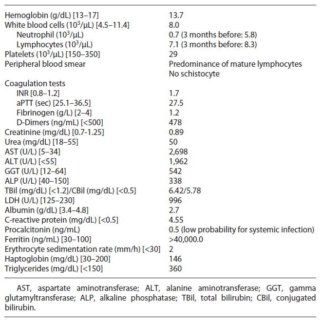
Upon admission, on physical examination the patient was hemodynamically stable, febrile (40°C), frankly jaundiced and a non-painful hepatomegaly was detected; the remaining examination was unremarkable, with no signs of encephalopathy, ascites, chronic liver disease, active arthritis, mucocutaneous bleeding, cutaneous purpura, or enlarged lymph node. Initial blood investigations revealed bicytopenia, severe liver injury, and increased INR with normal remaining coagulation (shown in Table 1). Periph eral blood smear showed predominance of mature lymphocytes. Computed tomography chest-abdomen-pelvis excluded signs of malignancy, enlarged lymph nodes, chronic liver disease, or biliary obstruction.

Table 1. Laboratory workup 

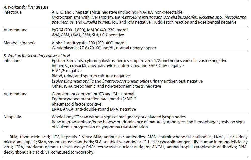
Initial septic workup (urine examination and imaging tests) ruled out infection; however pending the results of cultures, the patient was on antibiotic therapy. After 72 h, he remained febrile and developed organ failure: cardiovascular (systolic arterial pressure <90 mm Hg requiring inotropic support) and respiratory (PaO2/FiO2: 200-300 mm Hg requiring supplementary O2), so he was admitted to the intermediate care unit. Cultures have proven sterile, and extensive workup for liver disease was normal (shown in Table 2). Further workup revealed high ferritin, LDH, and triglycerides and low fibrinogen (shown in Table 1).

Table 2. Additional workup 

After excluding malignancy (normal computed tomography), infections (negative cultures and normal wide virus panel), and rheumatic disorder flare (no arthritis and normal complement, autoimmunity, and erythrocyte sediment rate), we considered HLH diagnosis attending to the cytopenias, hyperferritinemia, and persistent fever. An HLH score of 277 supported the diagnosis. Attending to organ failure, while we were waiting for the results of the bone marrow aspirate, we started dexamethasone (10 mg/m2 daily) and first dosage of etoposide (150 mg/m2). Meanwhile, the bone marrow aspirate showed hemophagocytosis, predominance of mature clonal lymphocytes, and no signs of progression nor transformation to aggressive lymphoma. The patient continued treatment with etoposide (150 mg/m2 biweekly for 2 weeks, then weekly) and dexamethasone (10 mg/m2 daily for 2 weeks with progressive tapering).

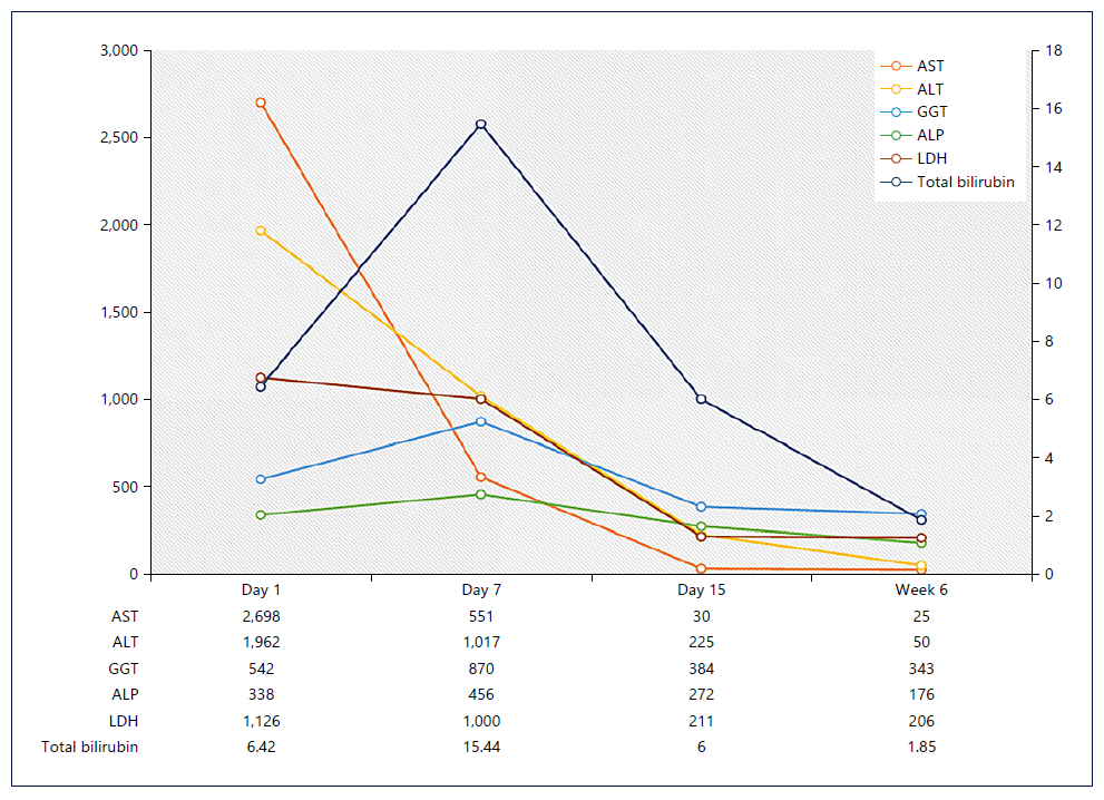
Rapid clinical (apyrexia and reversal of all organ failures) and liver improvement was observed (shown in Fig. 1), as well as neutrophil count and INR normalization. However, after 6 weeks of therapy, the patient developed febrile neutropenia related to immunomodulatory therapy, so we discontinued etoposide and started granulocyte colony-stimulating factor and broad-spectrum antibiotic therapy. The patient remained neutropenic and acquired a healthcare-associated pneumonia requiring intensive care admission, there was a progressive worsening, and he died 8 weeks after admission.

Fig. 1. Analytical evolution. 

Discussion

HLH results from uncontrolled activation of macrophages, natural killer cells, and T cells due to enhanced antigen presentation [3, 4]. This activation produces massive secretion of proinflammatory cytokines, a socalled cytokine storm, that directly contributes to end-organ damage and rapidly progressive multiorgan dysfunction [4].

The etiology of HLH is classified into primary (genetic) and secondary (reactive). In children, congenital defects in cytotoxic T cell and natural killer cell function and inflammasome dysregulation are described. Secondary causes result from acquired immune dysfunction in response to infections, malignancies, autoimmune diseases, or other causes (e.g., drugs, vaccination, organ/stem cell transplantation) [3, 6]. The underlying cause cannot be identified in 20% of cases [6, 7].

CLL is a monoclonal lymphoproliferative disease that results from the proliferation and accumulation of morphologically mature but immunologically dysfunctional B-cell lymphocytes [8]. HLH in the context of CLL has rarely been reported, mostly due to chemotherapy and CLL progression/transformation [9]. We emphasize that our patient had no evidence of progression of CLL or transformation into a more aggressive lymphoma. In CLL, there is a continuous crosstalk between dysfunc tional B and T lymphocytes [10, 11]. Disturbances in apoptosis of T cells, altered patterns of surface molecules of T cells, and unbalanced cytokine environment (IL-10, IL-6, IL-4) were described in CLL [10], which might contribute to uncontrolled activation of the immune system. Secondary HLH may be induced by autoimmune disorders, known as macrophage activation syndrome. Although our patient had rheumatoid arthritis, there were no arthritis nor analytical alteration suggesting flare.

Few case reports have described HLH following administration of vaccines [7, 12-14] including after COVID-19 vaccination [15-17], mostly in children with in-herited variants of HLH-associated genes or immunosuppressed adults. We postulated that the uncontrolled activation of the immune system resulted from an immune response triggered by vaccination (time frame) and an underlying immune defect (immunosuppression, malignancy). Dramatic proinflammatory responses have been identified in some individuals following immunization [18].

The clinical presentation of HLH is mostly non-specific with an acute or subacute course [3]. The liver is one of the most affected organs. ALT and AST elevations are seen in ∼85% of adult, mostly mild, and half of patients have hyperbilirubinemia [6, 7]. ALI/ALF as presentation is rare and usually occurs with concomitant organ failure [19]. Liver histopathological features include sinusoidal dilatation and hepatocellular necrosis [20]. Underlying liver disease, infiltration of activated histiocytes and over-production of cytokines are the putative mechanisms of liver injury [21].

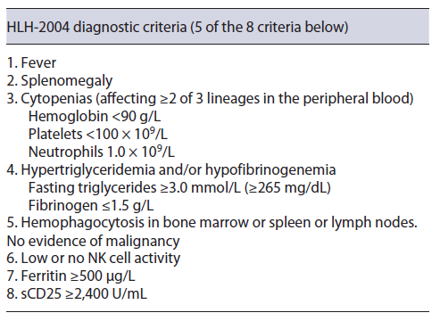
Pulmonary (42%), cutaneous (25%), and neurological (25%) involvement are also frequent [3, 22]. To make the diagnosis of HLH, the patient should meet five of the eight diagnostic HLH-2004 criteria (shown in Table 3) [23].

Table 3. HLH-2004 diagnostic criteria 

Cytopenias are the key laboratory markers of HLH. Hyperferritinemia is the finding that most often (90%) leads to suspicion of LHH [3, 23]. Hypertriglyceridemia or hypofibrinogenemia is easily determined. Although determination of soluble CD25 is helpful for diagnosis, it is rarely available [23]. Finally, the demonstration of hemophagocytosis in bone marrow is helpful for diagnosis, being also mandatory to exclude underlying malignant disorders [23]. In our patient, beyond the signs of hemophagocytosis in the bone marrow, there was no evidence of CLL progression nor transformation to aggressive lymphoma, so five criteria were met.

The HScore [24], which includes clinical and laboratory parameters, supports the diagnosis. Our patient predicted HLH with 99% of probability, so we started therapy immediately, given the presence of organ failure. The diagnosis of severe liver dysfunction induced by HLH is challenging, particularly in the early phase of the disease, as the presentation is non-specific, making it difficult to distinguish it other causes of ALI [3].

The prognosis of adult HLH is poor, with mortality rates ranging from 41 to 75% [3, 6, 19]. A delayed diagnosis is the limiting step toward a successful outcome, including hyperinflammation control (corticosteroids and etoposide) and treatment of the underlying cause, based on HLH-2004 protocols [3]. Early suspicion supported by a high HScore allowed the timely institution of successful treatment. The patient’s outcome was conse-quence of neutropenia and respiratory infection. Some authors recommend prophylactic antibiotic and antifungal therapy and granulocyte colony-stimulating factor in patients treated with T-cell depleting therapy [25], which was not started as early as desirable in our patient. Additionally, there is growing evidence of polyvalent immunoglobulin in HLH treatment [23], specially in our patient, as CLL is associated with lower humoral immunity and hypogammaglobulinemia.

HLH requires a high index of suspicion and should be considered in patients with unknown cause of severe liver dysfunction accompanied by sudden unexplained on set of systemic inflammatory response and multiple organ involvement. We alert to the need to monitor suspicious symptoms after vaccination (e.g., high fever) in patients with underlying pathology. The diagnosis of HLH should not be based on the fulfilment of criteria alone, and it is of utmost importance that an experienced hematologist judges the clinical aspects and weighs up the risks and benefits of treatment.

References

1. European Association for the Study of the Liver Electronic address easloffice@easlofficeeu; Clinical practice guidelines panel; Wendon J; Panel Members; Cordoba J, Dhawan A, et al. EASL Clinical Practical Guidelines on the management of acute (fulminant) liver failure. J Hepatol. 2017 May; 66(5): 1047- 81 [ Links ]

2. Breu AC, Patwardhan VR, Nayor J, Ringwala JN, Devore ZG, Ganatra RB, et al. A multicenter study into causes of severe acute liver injury. Clin Gastroenterol Hepatol. 2019 May; 17(6): 1201-3. [ Links ]

3. Ramos-Casals M, Brito-Zerón P, López-Guillermo A, Khamashta MA, Bosch X. Adult haemophagocytic syndrome. Lancet. 2014 Apr; 383(9927): 1503-16. [ Links ]

4. Aricò M, Danesino C, Pende D, Moretta L. Pathogenesis of haemophagocytic lymphohistiocytosis: review. Br J Haematol. 2001 Sep; 114(4): 761-9. [ Links ]

5. Allen CE, McClain KL. Pathophysiology and epidemiology of hemophagocytic lymphohistiocytosis. Hematology. 2015 Dec 5; 2015(1):177-82. [ Links ]

6. Li J, Wang Q, Zheng W, Ma J, Zhang W, Wang W, et al. Hemophagocytic lymphohistiocytosis: clinical analysis of 103 adult patients. Medicine. 2014 Mar; 93(2): 100-5. [ Links ]

7. Otrock ZK, Eby CS. Clinical characteristics, prognostic factors, and outcomes of adult patients with hemophagocytic lymphohistiocytosis: hemophagocytic lymphohistiocytosis of Adults. Am J Hematol. 2015 Mar; 90(3): 220-4. [ Links ]

8. Eichhorst B, Robak T, Montserrat E, Ghia P, Niemann CU, Kater AP, et al. Chronic lymphocytic leukaemia: ESMO Clinical Practice Guidelines for diagnosis, treatment and follow-up. Ann Oncol. 2021 Jan; 32(1): 23-33. [ Links ]

9. Kilari D, Venci N, Friedberg J, Bennett JM. Hemophagocytic lymphohistiocytosis masquerading as progressive chronic lymphocytic leukemia. Leuk Res Rep. 2013; 2(1): 4-6. [ Links ]

10. Vlachonikola E, Stamatopoulos K, Chatzidimitriou A. T cells in chronic lymphocytic leukemia: a two-edged sword. Front Immunol. 2020; 11: 612244. [ Links ]

11. Riches JC, Ramsay AG, Gribben JG. T-cell function in chronic lymphocytic leukaemia. Semin Cancer Biol. 2010 Dec; 20(6): 431-8. [ Links ]

12. Yahya AM, Al-Hammadi S, AlHashaykeh NO, Alkaabi SS, Elomami AS, AlMulla AA, et al. Case report: reactive lymphohistiocytic proliferation in infant with a novel nonsense variant of IL2RG who received BCG vaccine. Front Pediatr. 2021 Nov 2; 9: 713924. [ Links ]

13. Ikebe T, Takata H, Sasaki H, Miyazaki Y, Ohtsuka E, Saburi Y, et al. Hemophagocytic lymphohistiocytosis following influenza vaccination in a patient with aplastic anemia undergoing allogeneic bone marrow stem cell transplantation. Int J Hematol. 2017 Apr; 105(4): 389-91. [ Links ]

14. Lagousi T, Korovessi P, Panagouli E, Tsagris V, Kostaridou S. A rare case of measles-associated hemophagocytic lymphohistiocytosis in an infant. Cureus. 2020 May 23; 12(5): e8246. [ Links ]

15. Ai S, Awford A, Roncolato F. Hemophagocytic lymphohistiocytosis following ChAdOx1 nCov-19 vaccination. J Med Virol. 2022 Jan; 94(1): 14-6. [ Links ]

16. Attwell L, Zaw T, McCormick J, Marks J, Mc-Carthy H. Haemophagocytic lymphohistiocytosis after ChAdOx1 nCoV-19 vaccination. J Clin Pathol. 2022; 75(4): 282-4. [ Links ]

17. Tang LV, Hu Y. Hemophagocytic lymphohistiocytosis after COVID-19 vaccination. J Hematol Oncol. 2021 Dec; 14(1): 87. [ Links ]

18. Scully M, Singh D, Lown R, Poles A, Solomon T, Levi M, et al. Pathologic antibodies to platelet factor 4 after ChAdOx1 nCoV-19 vaccination. N Engl J Med. 2021 Jun 10; 384(23): 2202-11. [ Links ]

19. Lin S, Li Y, Long J, Liu Q, Yang F, He Y. Acute liver failure caused by hemophagocytic lymphohistiocytosis in adults: a case report and review of the literature. Medicine. 2016 Nov; 95(47): e5431. [ Links ]

20. Favara BE. Histopathology of the liver in histiocytosis syndromes. Pediatr Pathol Lab Med. 1996 Jan; 16(3): 413-33. [ Links ]

21. Chen JH, Fleming MD, Pinkus GS, Pinkus JL, Nichols KE, Mo JQ, et al. Pathology of the liver in familial hemophagocytic lymphohistiocytosis. Am J Surg Pathol. 2010 Jun; 34(6): 852-67. [ Links ]

22. Tseng YT, Sheng WH, Lin BH, Lin CW, Wang JT, Chen YC, et al. Causes, clinical symptoms, and outcomes of infectious diseases associated with hemophagocytic lymphohistiocytosis in Taiwanese adults. J Microbiol Immunol Infect. 2011 Jun; 44(3): 191-7. [ Links ]

23. La Rosée P, Horne A, Hines M, von Bahr Greenwood T, Machowicz R, Berliner N, et al. Recommendations for the management of hemophagocytic lymphohistiocytosis in adults. Blood. 2019 Jun 6; 133(23): 2465-77. [ Links ]

24. Fardet L, Galicier L, Lambotte O, Marzac C, Aumont C, Chahwan D, et al. Development and validation of the HScore, a score for the diagnosis of reactive hemophagocytic syndrome: score for reactive hemophagocytic syndrome. Arthritis Rheumatol. 2014 Sep; 66(9): 2613-20. [ Links ]

25. Kim YR, Kim DY. Current status of the diagnosis and treatment of hemophagocytic lymphohistiocytosis in adults. Blood Res. 2021 Apr 30; 56(S1): S17-25 [ Links ]

1Statement of Ethics Ethical approval was not required for this study, in accordance with local/national guidelines. Written informed consent was obtained from the patient’s next-of-kin for publication of this case report and any accompanying images.

3Funding Sources There are no funding sources for this case report.

5Data Availability Statement All data generated or analyzed during this study are included in this article. Further inquiries can be directed to the corresponding author.

Received: October 22, 2022; Accepted: January 12, 2023

Correspondence to: Cristiana Sequeira, c.afonso.sequeira@gmail.com

Conflict of Interest Statement The authors have no conflicts of interest to declare.

Author Contributions Material preparation, data collection, and first draft of the manuscript were performed by Cristiana Sequeira. Cristiana Sequeira, Sara Ramos Lopes, Anabela Neves, Inês Costa Santos, Cláudio Martins, and Ana Paula Oliveira read and approved the final manuscript.

Creative Commons License This is an open-access article distributed under the terms of the Creative Commons Attribution License