


 Inglês (pdf)
 Artigo em XML
 Referências do artigo
 Enviar este artigo por email
 Citado por SciELO
  Similares em
SciELO
Similares em
SciELO 
 Permalink
Introduction
Onychocryptosis or unguis incarnatus, commonly known as “ingrown nail”, is an inflammatory disease of the lateral nail fold originated by the embedding of the lateral border of the nail plate into the lateral sulcus1,2. This condition, usually presents as tenderness and edema and occurs most frequently in toes, particularly the hallux, is most frequent among young adult males, and has a high incidence estimated at 10,000 cases per year in the UK alone1-8.
Precipitating factors for onychocryptosis include extrinsic factors, such as improper (convex and deep) cutting of the ungual plate, tight and high-heeled footwear, trauma, bad hygiene, practice of sports requiring short sprints and stops, and intrinsic factors, as hallux valgus and other toe deformities, hyperhidrosis, obesity, type I diabetes, and subungual tumors, among others2,6,9-11. The main modifiable precipitating factor is convex cutting of the ungual plate, which leaves small splinters that penetrate the epidermis and trigger ungual ingrowth as the plate grows. Therefore, appropriate, straight, and shallow nail cutting is paramount in the prevention of onychocryptosis8.
Onychocryptosis can be broadly classified as mild, moderate, or severe and its treatment can be divided into conservative and surgical approaches, but both classification and preferred treatment options vary greatly in the reviewed literature7,12. In early and mild presentations conservative treatment is recommended, whereas in recurrences and more severe cases definitive surgical treatment is indicated7,12-14. In most cases, surgical approaches involve lateral horn matricectomy in order to narrow the ungual plate3,13,15-17. This matricectomy can be performed using different techniques, either chemical (88% phenol, sodium hydroxide 10 or 20% or trichloroacetic acid 80–100%) or surgical (excision, curettage, electrocoagulation, or laser ablation)3,9,13,18. In other cases, nail ingrowth is associated with considerable ungual lateral fold hypertrophy, and excision of part of peri-ungual soft tissues is a more adequate approach13. Several surgical alternatives are available, such as the Howard-Dubois technique, the Vandenbos technique, the Noël technique, the Tweedie and Ranger rotational flap, the Super U technique, and the Syme's terminal surgery3,18.
Ungual organ surgery, a specific branch of dermatologic surgery, is not broadly practiced and many specialists are uncomfortable performing it19. Therefore, it is essential to characterize in a single paper each and every of the presented therapeutic options, as well as their indications, technical difficulty, post-operative discomfort, aesthetic result, and recurrence rates.
The objective of this paper is to systematically review the relevant literature on surgical treatment of onychocryptosis in order to describe and compare the available surgical methods and their relevance in current clinical practice.
Methods
Clinical studies, review articles, and case reports were consulted in the PubMed and Clinical Key databases. The keywords chosen for the research were “onychocryptosis”, “ingrown nail” and “nail surgery.” The search was restricted to the English, French, Spanish, and Portuguese languages and extended to include the last 15 years of relevant publications due to the scarcity of literature fitting the search criteria. The bibliography was later further complemented by earlier references.
bibliographical references were analyzed and selected according to their relevance. Although the main scope was surgical treatment, we included some publications on epidemiology, physiopathology, classification, and conservative treatment to obtain a comprehensive review of the disease.
The present literature review was elaborated based on the research and its critical analysis.
Results
Staging
Classification of onychocryptosis based on severity is paramount to define the best therapeutic approach. However, several of these classifications exist, with none being unanimously used in clinical practice. Heifetz defined a three-stage classification, later reviewed by Mozena, who subdivided stage II into IIa and IIb8,12,14,20,21. Recently, this classification was expanded by Martinez-Nova et al., who added a fourth stage (Table 1)12,16,21.
Table 1 Onychocryptosis staging according to Martinez-Nova et al.21
| Stage I | Erythema, slight edema, and tenderness to pressure; lateral ungual fold does not surpass ungual plate limits |
|---|---|
| Stage II | Pain, erythema, and greater edema; hyperesthesia; lateral ungual plate abscess surpassing ungual plate limits. · IIa: Nail fold exceeds the nail plate <3 mm · IIb: Nail fold exceeds the nail plate >3 mm |
| Stage III | Severe symptoms; presence of granulation tissue; chronic hypertrophy of lateral ungual plate |
| Stage IV | Chronic deformity of ungual organ, including plate, lateral folds, and distal fold |
Conservative treatment
Dermatologists defending a non-invasive approach consider that the pathogenesis of onychocryptosis is preventable through the protection of lateral ungual sulcus against penetration by the ungual plate11. The first step for any conservative method is attempting to separate and cut the portion of ungual plate penetrating the lateral ungual fold6.
This low risk and low-cost strategy should be used alone only in mild to moderate cases, and several methods can be associated to obtain higher therapeutic success11,22. The literature is not consensual in defining the stage from which more invasive therapies are necessary8.
A good patient–doctor relationship, that capacitates the patient to perform his own treatment, and high compliance are essential to the success of conservative treatment and avoidance of recurrence, as the patient remains predisposed to onychocryptosis due to the imbalance between ungual plate wideness and nail bed11.
Preventive measures such as choice of appropriate shoe wear and straight and shallow cutting of the nail plate should be adopted permanently by all patients2,15.
TAPING
Applying tape to the lateral ungual fold, to exert traction in an oblique proximal direction and separate it from the ungual plate, is the least aggressive method. When performed frequently and correctly, it can resolve mild cases. It is less effective in patients who sweat abundantly and whenever the onychocryptosis is in advanced stages, as the presence of purulent drainage displaces the tape8,9,23.
PACKING
Consists in the insertion of small, antiseptic-embedded cotton between the corner of the ungual plate and the ungual fold. This procedure should be repeated daily, attempting a progressive separation of structures with larger portions of cotton. Results are good for stage I patients, however, it requires a long treatment duration to be effective. Dental floss can be alternatively used7,9,15,17.
GUTTER TECHNIQUE
A more invasive approach consists in applying a plastic gutter to cover the ungual lateral border. This gutter can be constructed from the longitudinal cut of an intravenous fluid tube or other sterile plastic tubing. The border of the ungual plate is separated from the sulcus and ungual folds and the gutter is inserted alongside it, not allowing it to penetrate the surrounding soft tissues. The gutter is fixed to tape, acrylic glue, or sutures and is removed 6–8 weeks later, during which the inflammation subsides (Fig. 1)8,9,19,24.
UNGUAL ORTHOSES
Onycho-orthoses were developed to treat cases in which the onychocryptosis is mainly due to a marked curvature of the ungual plate, which can result in pincer nails or unguis constringuens, with marked recurrent pain and high morbidity. These are ungual traction devices that attempt to decrease plate curvature.
Several types are available. One of the most used consists of a steel wire applied in the dorsal side and hooked to both borders of the ungual plate. Alternatively, plastic bands or super elastic tape can be used. It is necessary that the wire material has memory, so that it causes continuous traction over the plate, making it increasingly flat. Nail braces are one of podiatrists favorite methods3,14.
In 2015, Güler et al.14 conducted a retrospective study which compared ungual orthoses placement with Winograd wedge excision on 159 patients, diving it between two groups, group I, with 74 subjects, treated with ungual orthoses, and group II, with 85 patients, subjected to Winograd technique. The study reported a shorter recovery time with a quicker return to work in group I (4.15 ± 1.07 days; p < 0.001) as well as higher patient satisfaction (94.6% vs. 82.4%; p < 0.05). No statistically significant differences were reported with regards to recurrence rates, (8.1 and 9.4% respectively for groups I and II) with a follow up of less than two years.
A prospective study was conducted by Erdogan et al.25 in 2014, which reported a recurrence rate of 12.4% in 88 patients with onychocryptosis treated with onycho-orthoses. However, the study included patients with all severity stages, so we cannot infer on its utility in each stage based on this high recurrence rate.
This procedure is simple, cost-effective, safe (even in patients with comorbidities) and can be applied for long periods and reapplied in case of recurrence.14
INTRALESIONAL THERAPY
Intralesional injection of corticosteroids is an option that dermatologists are familiar with for other conditions. In 2019, Vílchez-Márquez et al.26 reported 5 cases of stages II and III onychocryptosis treated with intralesional injections of 0.5–1 mL of a 40 mg/mL commercial solution of triamcinolone acetonide, diluted at a ratio of 1:5 in an anesthetic solution of mepivacaine 2%. No relapse was observed during a minimum of 6 months follow-up of the patients26.
ANTIBIOTHERAPY
Although regularly prescribed, the use of oral antibiotics was not demonstrated to be as efficient as conservative treatment of onychocryptosis, even in cases with infection and granulation tissue9,17.
HYGIENIC MEASURES
An appropriate foot hygiene and avoidance of tight shoe wear is essential during conservative treatment in order to reduce inflammation and prevent infection7. Daily or twice daily washing with iodopovidone or potassium permanganate 1:10000 should be performed followed by topical antibiotic or medium to high power topical corticosteroid application. It is important to avoid trauma and promote straight and shallow cutting of the nail plate, leaving the distal lateral corner of the plate which should be superficially filed2,7,11,17.
Surgical treatment
On a first stage I episode, it is considered appropriate to conservatively prevent the ungual plate spike from penetrating the lateral ungual sulcus epidermis; however, for recurrences or stage II and higher presentations, surgical treatment is recommended16. This approach consists in partial avulsion of the ungual plate associated to a chemical or surgical measure to prevent recurrences17.
Isolated total avulsion of the ungual plate, although routinely performed in the past and still used by some clinicians, should be avoided, as it does not provide a definitive solution and even worsens the condition: the absence of ungual plate leads to the elevation of the distal finger pulp, forming a false distal wall in which the plate will penetrate when it grows again, leading to distal ingrowing9,11.
Surgical approaches aim to definitively correct onychocryptosis through permanent narrowing of the ungual plate achieved by matricectomy or soft periungual tissue resection. Recently, a retrospective study published in 2021 showed satisfactory results without the latter. A 36-month follow-up of 2118 patients showed a recurrence rate of 1.7% with only debridement of granulation and partial avulsion of the nail plate with scalpel or scissors. No sutures were used, neither electrocauterization27.
Partial avulsion presents recurrence rates of approximately 83%28, and so is no longer performed in isolation. Consequently, other strategies for definitive narrowing of the ungual plate are currently performed, such as matricectomy of the matrix portion corresponding to the area of removed plate6,12.
Surgical treatment - Matricectomy
CHEMICAL MATRICECTOMY
Chemical cauterization with 88% phenol solution was first described by Boll in 1945 and later popularized by Bell in 1977 and is currently the preferred technique among dermatologists6,23.
Phenol has simultaneously necrotizing, antiseptic and anesthetic properties which result in high indexes of success and patient satisfaction. The necrotizing effect induces ungual matrix destruction and consequently stagnates growth of the cauterized plate; the antiseptic effect allows surgery of moderately infected patients to be performed; the anesthetic effect, through demyelination of nerve terminals for a few weeks, increases post-operative patient comfort9,23.
After local anesthesia by digital block with 2% lidocaine without adrenaline and the application of a tourniquet, the granulation tissue, if present, is curetted to obtain a better visualization of the lateral border of the ungual plate, and avoid overextending the partial avulsion13. It is essential that the areas to be cauterized are free of hematic content since blood neutralizes phenol and is the main cause of technique failure. After partial avulsion, 3–5 mm wide, of the ungual plate, a cotton swab imbedded in phenol 88% is applied to the lateral horn of the matrix and the portion of exposed ungual bed, for 1–3 min, without dripping, and with care being taken to avoid cauterizing the bed under the remaining ungual plate or periungual tissues, as excessive cauterization may result in ungual dystrophy6,9,13,23. Postoperatively the limb should be elevated for 24 h, after which the band-aid is removed, and the wound washed with hydrogen peroxide 3%13. Analgesics are rarely necessary, and the patient should be evaluated one week after surgery6.
Phenolization is a simple and inexpensive technique, with reduced postoperative discomfort, quick return to daily activities, low rate of complications and recurrences, and good aesthetic results. The main disadvantage of its use is long-term postoperative exudation, lasting an average of 17 days, that might promote infections in patients lacking adequate hygiene. The patient must be instructed to wash the affected toe twice a day and avoid closed shoes until exudation ceases. Exudation time can be shortened with use of a 20% ferric chloride solution after phenolization or with the use of adrenaline in anesthesia. Postoperative follow up should be conducted in days 1, 10, 30, and 60 after intervention. This technique can be safely used in diabetic patients and those under anticoagulation13,29,30.
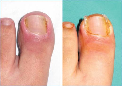
Chemical matricectomy is considered a low difficulty surgical technique and should be dominated by all dermatologists. It is indicated in patients with onychocryptosis stage I and IIa as well as pincer nails (Fig. 2)21,2 3,24.
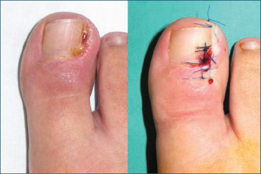
Figure 2 Partial avulsion followed by chemical matricectomy using 88% phenol solution. Source: Dermatology Surgery Unit – Coimbra University Hospital.
Di Chiacchio et al.31 reported, in 2010, for a follow up of 33 months, a recurrence rate of 1,9% in 267 patients with onychocryptosis subjected to this technique, findings concurring with those of Kimata et al.32 (1.1%); Bostanci et al.33 (0.6%) and Andreassi et al.34 (4.3%). The aforementioned studies compound to a total of 2102 cases, with similar results; consequently, we can state that phenolization of ungual matrix yields excellent results, cosmetically and functionally, with very low recurrence rates13. More recently, a 5 year retrospective study of 520 patients treated with classic surgical matricectomy and chemical matricectomy with phenol, showed a higher recurrence rate with phenolization (17.2% vs. 8.2%), but a shorter postoperative period, decreased intensity and duration of pain, a lower risk for surgical wound infection, and improved cosmetic outcomes16.
Instead of phenol, sodium hydroxide 10% or trichloroacetic acid 80–100% can be used. The former decreases mean time of exudation to 9 days and can be applied for only 1 min, while the latter results in fast healing of approximately 2 weeks3,9,18,23.
SURGICAL MATRICECTOMY BY CURETTAGE OR ELECTROCOAGULATION
After partial avulsion of the ungual plate, the ungual matrix is excised with a scalpel, followed by curettage or electrocoagulation. It may be necessary to incise the skin in the most medial projection of the matrix to be removed in order to facilitate visualization of the matrix and guarantee that no remnants of it are left behind. A surgical microscope can be used35.
Kim et al.36 conducted a prospective study analyzing this technique, comparing matricectomy by curettage and by electrocauterization, after partial avulsion, on a sample of 62 patients. The rate of infection was 15.6% and 10.3% and recurrence rate was 6.2% and 13.8% on patients submitted to curettage and electrocoagulation, respectively. This study concluded that curettage is a viable alternative in military field hospitals and some primary care institutions where medical supplies are limited.
In electrocoagulation, the large amount of heat applied may lead to thermal periostitis, with long-standing postoperative pain.
Both techniques are best avoided as first line therapy, since they yield more complications and recurrences than other more efficient methods, such as phenolization.
Wedge excisions are another type of surgical matricectomy, but they will be reviewed separately due to their complexity and simultaneous resection of periungual soft tissues.
LASER OR RADIOSURGERY MATRICECTOMY
In these similar techniques, matricectomy is performed after partial avulsion of the ungual plate identical to that performed in chemical matricectomy.
Concerning laser matricectomy, CO2 laser is the most used, and the matrix and ungual bed are vaporized by a laser beam with power and size varying according to the user's expertise and equipment used. Its main advantage is its hemostatic effects. Experienced surgeons can reach high success rates, similar to those of chemical matricectomy, however, this procedure is considerably expensive19,21,23,24.
In radiosurgery, the electrode remains cold during the procedure, which allows for very selective matrix ablation. After partial avulsion, the malleable, sword-shaped electrode, is inserted horizontally between the matrix and the proximal ungual fold. Its dorsal face is coated by a material that avoids damage to the proximal nail fold, usually Teflon. The power and duration of ablation depend on the equipment used. Radiosurgery yields excellent results but is quite onerous17,21,24.
Both methods achieve a very low recurrence rate (< 5%) and cosmetic results, but large-scale comparative studies are not available and high costs may prevent its widespread application9,18,23,24.
WEDGE EXCISION
This type of surgical procedure involves a bulk resection of a strip of lateral longitudinal ungual plate with the corresponding matrix and ungual bed, together with a portion of lateral ungual fold and granulation tissue. The Winograd technique is the most known of these techniques, but other alternatives are available, such as the Zadik or Emmert procedures or the knot technique23,37.
After local anesthesia and application of an elastic tourniquet, a vertical incision is made on the plate and ungual bed, 2–3 mm from the border on the affected side, starting at the most proximal matrix projection, sliding the scalpel longitudinally and distally. Another incision, with the same start and end points, is made along the fold, excising a part of it, forming a wedge-shaped piece. The lateral horn of the remaining matrix is exposed and removed with the blade or by curettage, and the wound is closed by direct suture6,38.
The main disadvantage of this technique is postoperative pain due to trauma caused in the periosteum during matrix resection; secondary infections are also reported in around 20% of patients. The success rate is highly dependent on surgeon experience, and recurrence is more operator-dependent compared to that of more simple techniques, such as chemical cauterization23.
A case series published in 2014 by Guler et al.39 reported, in a 239 sample of patients subjected to the Winograd procedure, a 96.6% satisfaction rate with cosmetic results. However, in 2011, Kayalar et al.40 reported a recurrence rate of 9.8% on a cohort of 224 patients, with 7.1% needing surgical revision.
Novel technical modifications have been described. Recently, Uygur et al.38 tested a different suture technique through a prospective study comparing it to the traditional suture technique after Winograd procedure. The results showed a recurrence rate of 14.1% for patients randomly assigned the new suture, compared to 33,3% in the traditional method, for a 12-month follow up.
Independently of the technique used, wedge excisions are associated with high recurrence rates, weak aesthetical and functional results, and significant morbidity relatively to other techniques9.
Surgical treatment - periungual soft tissue resection
Onychocryptosis, if untreated, worsens through soft periungual tissue hypertrophy, which is unaesthetic and prevents healing through conservative methods. In the most serious cases, hypertrophy extends distally, with a false ungual distal fold forming. In these cases, even surgical treatments to definitely narrow the ungual plate may be insufficient, and soft periungual tissue resection may be necessary if hypertrophy is significant18,23.
HOWARD-DUBOIS PROCEDURE
This procedure is indicated in cases of ingrowth of the distal border of ungual plate and in some cases of moderate to severe lateral ungual fold hypertrophy13,23. After cleaning with alcohol 70%, tourniquet application and anesthesia by digital blockage with lidocaine 2%, a watermelon-slice shaped portion of tissue is excised parallel to the hyponychium, approximately 5 mm below the distal sulcus and measuring 3–7 mm at the thickest point, around the fingertip. Tissue is removed until the distal phalanx plane is reached, after which the borders are sutured with interrupted 4'0 or 5'0 sutures and the hyponychium is pulled downwards, eliminating the false distal ungual fold. The lateral ungual folds are also pulled, mainly in their union with the lateral sulcus, where ungual ingrowth is more common9. A rare, but possible complication is necrosis of the margins if sutures are too tight. This procedure is relatively invasive and causes mild to moderate postoperative pain, and analgesics should be prescribed. The limb should be elevated for 48 h and the dressing changed after the first 24 h. The sutures are removed after 14 days (Fig. 3)13.
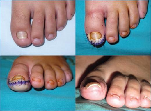
Figure 3 Howard-Dubois surgical procedure. Moderate hypertrophy of lateral and distal nail folds without granulation tissue (upleft). Post-surgical result in upper view (upright) and anterior view (downleft). Final result after healing (downright). Source: Dermatology Surgery Unit – Coimbra University Hospital.
The results of this technique are described by Di Chiacchio and Di Chiacchio13, with excellent cosmetic outcomes. However, no studies concerning efficacy were found.
VANDENBOS PROCEDURE
The Vandenbos procedure is indicated in cases of severe hypertrophy of the lateral ungual fold that cover a significant part of the ungual plate.
This procedure involves ample resection of periungual tissue without avulsion of the ungual plate or matrix. After disinfection, anesthesia is achieved by digital block with lidocaine 2% or bupivacaine 0.5% and a tourniquet is applied. An incision is made along the lateral ungual sulcus, from the distal to the proximal side, up to the junction of the lateral and proximal ungual folds. A second incision starts at the end of the first and extends laterally to the inferior third of the finger18,20,22.
The resection yields a 1,5 × 3 cm defect, with a portion of distal phalanx sometimes exposed. Hemorrhage is controlled through electrocauterization of silver nitrate appliance. This defect heals by second intention in a period from 4 to 6 weeks18,20,22.
The advantages of this technique are its easy execution, low levels of postoperative pain, and minimal risk of ungual plate dystrophy since the procedure does not involve the matrix. The longer recovery time is compensated by its excellent aesthetical and functional results in the medium to long term9,20.
A retrospective study by Chapeskie and Kovak22 in 2010, relative to the application of the Vandenbos technique to 212 patients, found no recurrences and excellent aesthetical result in all cases after a follow up of 8 years.
NAILTEST20, a non-randomized study published in 2017, analyzed complications and pain, function, and life quality in 39 patients from 4 to 20 years old and 59 ungual organs subjected to Vandenbos, and no recurrences were reported. Patient follow up visits were conducted at 1, 2 and 6 months postoperatively. 7 patients (18%) reported minor complications, namely hemorrhage (8%), excessive pain (8%), and infection (2%). The study yielded excellent functional results, low rate of complications, fast recovery time (median labor absenteeism was 7 days), and high patient satisfaction indexes (95%).
SUPER U
This technique, developed by Perez Rosa, is similar but more invasive compared to Vandenbos, comprising the removal of all excess periungual tissue in a U-shaped band. This involves the excision of both the lateral ungual folds as well as the distal fold. Another difference to the Vandenbos technique is that hemostasis is achieved through suture of the incision borders rather than cauterization. Healing occurs through second intention. Super U is indicated in cases of severe hypertrophy of both the lateral and distal folds9,41.
After anesthesia by digital block and tourniquet placement, a relatively horizontal U-shaped incision is made from the proximal portion of one of the lateral folds to the opposite proximal lateral fold13. Another incision is initiated at the end of the first, and runs through the lateral sulcus, distal sulcus (formed due to hypertrophy of the adjacent tissues, creating a false ungual distal fold) and the opposite lateral sulcus, ending the start of the first incision. A running locked, 3 or 4'0 non-absorbable suture is performed around the first incision (Fig. 4). The affected limb should be elevated for 48 h and strong analgesics such as tramadol should be prescribed for severe postoperative pain. Dressing should be removed by the surgeon 2 days after surgery and the finger washed with 3% peroxide hydrogen. Dressings should be switched every two days and rinsed in antiseptic ointment; closed shoe wear should be avoided until healing is complete13.
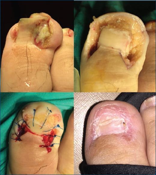
Figure 4 Super U technique. Severe hypertrophy of lateral and distal nail folds with abundant granulation tissue, purulent discharge and chronic deformity of ungual organ (upleft). After excision of granulation tissue and lateral and distal nail folds (upright). After suture the remaining borders (downleft). Final result after healing (downright). Source: Dermatology Surgery Unit – Coimbra University Hospital.
Infections are rare if appropriate hygiene is administered and should be treated with antibiotics. The long recovery time, about 2 months, is the main disadvantage of this technique, but symptomatic and functional improvement is dramatic when the Super U technique is applied to severe cases13.
In 2017, Correa et al.42 reported 2 recurrences in a 10-patient retrospective study. These 2 cases occurred in onychocryptosis with 4 and 6 years of evolution. The authors concluded the technique is very useful in severe cases of onychocryptosis with lateral and distal fold hypertrophy.
NOËL PROCEDURE
Two wedge shaped blocks of periungual tissue adjacent to the ungual fold are excised vertically on both sides of the plate, including fibrotic and granulation tissue. Depth of resection must be sufficient to involve the two superior thirds of the finger. The plate, ungual bed, and matrix are not involved in the resection and the defect is sutured with 4'0 interrupted sutures9.
This procedure can be performed only on one side of the ungual organ, but requires significant surgeon experience, as the location of one of the incisions close to the distal phalanx makes it difficult to execute.
Noël43, in a 23 patient-study, reported no recurrences after 1 year of follow up, as well as excellent cosmetic results. No other studies were found reporting the use of this technique.
TWEEDIE AND RANGER ROTATIONAL FLAP
This procedure consists in creating a flap of lateral periungual tissue that is transposed to a more inferior position. After curettage of the granulation tissue, a vertical slice of the ungual fold's distal base is excised, leaving a loose portion of tissue that is sutured.
The original study published by Tweedie and Ranger44 reported a success rate of 92%, but no other papers analyzing this technique were found.
KNOT TECHNIQUE
Presented in 2015 by Ince et al.45, the knot technique consists in a wedge excision including only periungual soft tissues without interfering with the ungual plate or bed. The excision is made in the area most frequently jammed against the plate, with the distal portion of the lateral ungual fold being removed and the suture performed so that the lateral distal plate corner lies superficial to the periungual tissues. The study had an average follow up of 20 months on 30 patients (34 procedures on 18 stage II and 16 stage III onychocryptosis) and reported no post-operative infection and just one instance of recurrence.
Later in 2015, Ince et al. conducted a prospective study comparing the Winograd procedure with the knot technique with the latter yielding a lower recurrence rate (2.2% vs. 17.7%).
Although results were excellent, additional, larger, and more representative studies are needed, as well as comparative trials with more efficient methods than Winograd serving as control, to access accurately the future role of this technique in the treatment of onychocryptosis.
Surgical treatment - Syme's terminal surgery
Consists in amputating the distal portion of the affected finger, involving avulsion of the ungual plate, ungual bed, and matrix resection, amputation of the distal half of the distal phalanx and suture of the inferior flap with skin from the dorsal face of the finger, resulting in a small and bulbous finger. It is a mutilating surgery and should be considered as a last resort9.
Conclusions
The therapeutic approach of onychocryptosis includes a vast array of options, and it is challenging to select the most effective for each degree of severity. Two main approaches can be adopted, conservative and surgical.
Conservative treatment is effective for stage I and consists of patient education and techniques that separate the border of the ungual plate and lateral ungual fold, such as taping, packing, the gutter technique, and nail braces. The latter should be used whenever the plate has a high curvature.
Surgical treatment is more suited to stages II and III. According to the analyzed literature, the preferential option is matricectomy with 88% phenol, which permanently narrows the plate, effectively preventing recurrences. Other chemical agents such as sodium hydroxide 10% and trichloroacetic acid 80–100% can be used, as well as surgical matricectomy by curettage, electrocautery, radiation, laser ablation, or wedge excision.
In stages II and III, permanent narrowing of the ungual plate should be preferred; however, in cases in which ungual fold hypertrophy is present, healing is only achieved through periungual soft tissue resection. The Howard-Dubois technique is preferred in mild lateral and distal hypertrophy, whereas the Vandenbos technique is warranted in severe lateral hypertrophy, and the Super U in severe lateral and distal hypertrophy.
Further comparative studies and randomized clinical trials are necessary to outline a standardized approach to the treatment of onychocryptosis, as the current literature is scarce and often contradictory.

