SciELO - Scientific Electronic Library Online

 
vol.81 número1Angiossarcoma de couro cabeludo em paciente idoso: um relato de casoDesenvolvimento de Carcinoma Baso Celualr após o uso de Metformina – Potencial papel das Nitrosaminas como Fatores Favorecedores índice de autoresíndice de assuntosPesquisa de artigos
Home Pagelista alfabética de periódicos  

Serviços Personalizados

Journal

Artigo

Indicadores

Links relacionados

  • Não possue artigos similaresSimilares em SciELO

Compartilhar


Portuguese Journal of Dermatology and Venereology

versão impressa ISSN 2795-501Xversão On-line ISSN 2795-5001

Port J Dermatol Venereol. vol.81 no.1 Lisboa mar. 2023  Epub 09-Fev-2023

https://doi.org/10.24875/pjdv.22000024 

CASE REPORT

Nodular hidradenoma mimicking nodular basal cell carcinoma

Hidradenoma nodular simulando carcinoma basocelular nodular

1Department of Dermatology, Hospital Municipal Universitário de Taubaté (H.MUT/UNITAU)

2Department of Dermatology, Departamento de Medicina da Universidade de Taubaté, UNITAU

3Department of Pathological Anatomy, Hospital do Servidor Público Estadual de São Paulo. São Paulo, Brasil


Abstract

A 40-year-old woman, phototype III, presented with an erythematous, sessile nodule with small ulceration points on the right forearm for 1 year and 6 months. Dermoscopy showed structures similar to blue-gray globules, arboriform telangiectasias with some thickened vessels, bright white spots, and microulceration points. The main hypothesis of nodular basal cell carcinoma (BCC) and excisional biopsy of the lesion were performed. Histopathology showed nodular adnexal epithelial neoplasia without atypia, with a focal connection to the epidermis, a clear cell component, and immunohistochemistry positivity for p63, epithelial membrane antigen (EMA), and cytokeratin AE1/AE3, concluding the diagnosis of nodular hidradenoma. The unusual presentation, mimicking a nodular BCC, and the importance of the correct approach to atypical lesions motivated this report.

Keywords Acrospiroma; Basal cell Carcinoma; Dermoscopy; Histology; Immunohistochemistry

Resume

Mulher de 40 anos, fototipo III, apresentando nódulo eritematoso, séssil, com pequenos pontos de ulceração no antebraço direito há um ano e meio. À dermatoscopia visualizadas estruturas semelhantes a glóbulos azul-acinzentados, telangiectasias arboriformes com alguns vasos espessados, manchas branco-brilhantes e pontos de micro ulceração. Realizada hipótese principal de carcinoma basocelular (CBC) nodular e biópsia excisional da lesão. A histopatologia evidenciou neoplasia epitelial anexial nodular sem atipias, com conexão focal com a epiderme e componente de células claras e a imunohistoquímica positividade para p63, antígeno de membrana epitelial (EMA) e citoqueratina AE1/AE3, concluindo o diagnóstico de hidradenoma nodular. A apresentação incomum, simulando um CBC nodular e importância da abordagem correta de lesões atípicas motivaram este relato.

Palavras-chave Hidradenoma nodular; Carcinoma basocelular; Dermoscopia; Histologia; Imuno-histoquímica

Introduction

Nodular hidradenoma, first described by Mayer in 1941, is a benign adnexal neoplasm with eccrine or apocrine differentiation that occurs in middle-aged adults, mainly women, and with a controversial prevalence in the literature1-3. Clinically, it manifests as a solid or cystic nodule, normochromic to erythematous, with a smooth surface, slow and endophytic growing, and with rare cases of exophytic growing. The most common affected locations are the scalp, face, trunk, and proximal extremities1. At dermoscopy, the pattern consisting of a homogeneous bluish area that occupies the entire lesion, associated with vascular structures and white dots, is the most common pattern4. The unusual presentation, mimicking a nodular basal cell carcinoma (BCC) and the importance of the correct approach to atypical lesions motivated this report.

Clinical case

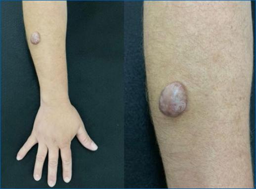
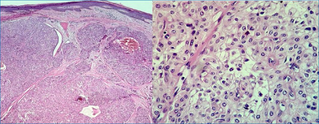
A 40-year-old female patient, phototype III, complained of a “lump” in her right forearm for a year and a half. Dermatological examination revealed a hyperchromic, erythematous, and sessile nodule (2.5 × 2 cm) of firm consistency and with points of ulceration (Figure 1). History of initial growth and subsequent stabilization, mild pain on manipulation. The diagnostic hypotheses of nodular BCC, dermatofibrosarcoma protuberans, amelanotic melanoma, Merkel cell carcinoma, and adnexal tumors were considered. On dermoscopy, the presence of structures similar to blue-gray globules, arboriform telangiectasias with some thickened vessels, bright white spots, and points of microulceration (Figure 2). An excisional biopsy was performed and the material was sent for histopathological and immunohistochemical examination. Histopathology showed nodular adnexal epithelial neoplasia without atypia, with a focal connection to the epidermis and a clear cell component, and characteristics suggestive of nodular hidradenoma (Figure 3); confirmed by immunohistochemistry with positivity for p63, epithelial membrane antigen (EMA), and cytokeratin AE1/AE3. The patient remains under follow-up with a good surgical scar aspect and no recurrence.

Figure 1 Hyperchromic, erythematous, sessile nodule of firm consistency, and with points of ulceration. 

Figure 2 Lesion dermoscopy. 

Figure 3 Nodular adnexal epithelial neoplasm without atypia, with a focal connection to the epidermis, and clear cell component (HE 40 and 400X). 

Discussion

Nodular hidradenoma is a benign adnexal neoplasm with eccrine or apocrine differentiation that occurs in middle-aged adults, mainly women1,2, as observed in this case. The exophytic clinical presentation is rare, however, described in the literature.1

There is considerable confusion in the literature regarding the appropriate nodular hidradenoma designation, and it has already been called clear cell hidradenoma, cystic nodule hidradenoma, clear cell myoepithelioma, and eccrine acrospiroma. This reflects different approaches among authors regarding its histological characteristics and histogenesis1,3.

The histopathology of nodular hidradenoma is characterized by the presence of a well-circumscribed but not encapsulated tumor. It presents a typical biphasic cell pattern, with polyhedral cells with eosinophilic cytoplasm and large cells with abundant clear cytoplasm and a small nucleus. There is a variable proportion between cell types, but clear cells predominate in one-third of cases1,2, as seen in this case. Immunohistochemistry shows reactivity for p63, EMA, and cytokeratin AE1/AE25, also observed in this case.

Classically, the differential diagnosis includes other adnexal tumors, being clinically indistinguishable1. Basal cell carcinoma (BCC) (ulcerated forms) as a differential diagnosis of hidradenoma nodular is rarely cited in the literature6.

Curative treatment consists of surgical excision, but the possibility of recurrence exists. Malignant transformation is rare1.

The present report registers an unusual presentation of a nodular hidradenoma, with hyperchromic, ulcerated, and exophytic manifestations, simulating a nodular BCC in the clinical aspect but mainly dermoscopically. We emphasize the importance of the correct approach to atypical lesions by the dermatologist, as well as the fundamental role of complementary exams (histopathology/immunohistochemistry) in the conclusive diagnosis of this case.

REFERENCES

1. Sousa VB, Lacerda VC, D'Elia MLN, Alves NTM, Pineiro-Maceira JM, Silva SCMC. Clear cell hidradenoma:atypical presentation on the scalp. Surg Cosmet Dermatol. 2016;8:S735. [ Links ]

2. Nandeesh BN, Rajalakshmi T. A study of histopathologic spectrum of nodular hidradenoma. Am J Dermatopathol. 2012;34:461–70. [ Links ]

3. Kazakov DV, McKee PH, Michal M, Kacerovska D. Lesions with predominant apocrine and eccrine differentiation. In:Dmitry V. Kazakov MD PhD, Cutaneous adnexal tumors. WKL. 2012;p1–171. [ Links ]

4. Serrano P, Lallas A, Del Pozo LJ, Karaarslan I, Medina C, Thomas L, et al. Dermoscopy of nodular hidradenoma, a great masquerader:a morphological study of 28 cases. Dermatol. 2016;232:78–82. [ Links ]

5. Canedo T, de Almeida MP, Cuzzi T, Ramos-e-Silva M. Immunophenotypic aspects of cylindroma and nodular hidradenoma. J Eur Acad Dermatol Venereol. 2010:24:178–85. [ Links ]

6. Montealegre Gómez CE, Correa Londoño LA. Hidradenoma de células claras:Presentación de un caso y revisión de la literatura. Rev Asoc Colomb Dermatol Cir Dematol. 1996;5:76–8. [ Links ]

FundingNone.

Ethical disclosures

Protection of human and animal subjects. The authors declare that no experiments were performed on humans or animals for this study.

Confidentiality of data. The authors declare that they have followed the protocols of their work center on the publication of patient data.

Right to privacy and informed consent. The authors have obtained the written informed consent of the patients or subjects mentioned in the article. The corresponding author is in possession of this document.

Received: September 26, 2022; Accepted: November 26, 2022

*Corresponding author: Flávia R. Ferreira E-mail: dermagica@uol.com.br

Conflicts of interest

None.

Creative Commons License Portuguese Society of Dermatology and Venereology. Published by Permanyer. This is an open access article under the CC BY-NC-ND license