SciELO - Scientific Electronic Library Online

 
vol.81 número2Toxidermia liquenóide associada ao esomeprazolUm caso de dermatose neutrofílica do dorso das mãos unilateral índice de autoresíndice de materiabúsqueda de artículos
Home Pagelista alfabética de revistas  

Servicios Personalizados

Revista

Articulo

Indicadores

Links relacionados

  • No hay articulos similaresSimilares en SciELO

Compartir


Portuguese Journal of Dermatology and Venereology

versión impresa ISSN 2795-501Xversión On-line ISSN 2795-5001

Port J Dermatol Venereol. vol.81 no.2 Lisboa jun. 2023  Epub 01-Mar-2023

https://doi.org/10.24875/pjdv.22000047 

CASE REPORT

Granular cell tumor: rare presentation in pediatric age

Tumor de células granulares: apresentação rara em idade pediátrica

1Department of Dermatology and Venereology, Centro Hospitalar e Universitário de São João (CHUSJ)

2Department of Pathological Anatomy, Centro Hospitalar e Universitário de São João (CHUSJ), Porto, Portugal


Abstract

Granular cell tumors (GCT) are rare soft tissue neoplasms that usually present as solitary lesions, although occasionally, they may be multiple. Most of these tumors arise in the oral mucosa and skin of adults, usually evolving as a slow-growing tumors. It is considered very rare in children. Malignant transformation is very rare, and only 2% of cases are known to spread to distant sites. Here we report a case of a 10-year-old child with a hard painless nodule on the tongue evolving for 6 months. Histopathological examination revealed infiltration of the tongue mucosa by polygonal cells with small nuclei and abundant pale eosinophilic granular cytoplasm expressing S100 protein strongly and diffusely, compatible with a granular cell tumor. A right partial glossectomy was performed. This case highlights the importance of mucosal biopsy for the diagnosis. It also emphasizes that regardless of pediatric age, a biopsy should not be postponed whenever there is uncertainty in the clinical diagnosis.

Keywords Granular cell tumor; Oral mucosa; Tongue

Resumo

Os tumores de células granulares são neoplasias raras dos tecidos moles e geralmente surgem como lesões solitárias, embora ocasionalmente sejam múltiplas. A maioria destes tumores ocorre na mucosa oral e na pele de adultos, geralmente evoluindo como um tumor de crescimento lento. Raramente surge em idade pediátrica. A transformação maligna é muito rara, e apenas 2% dos casos podem metastizar para locais distantes. Apresentamos o caso de uma criança de 10 anos com um nódulo duro, indolor, na língua com 6 meses de evolução. O exame histológico revelou infiltração da mucosa lingual por células poligonais exibindo núcleos pequenos e citoplasma granular, eosinofílico pálido, com expressão difusa de S100, compatível com tumor de células granulares. Foi realizada glossectomia parcial direita. Este caso salienta a importância da biópsia de mucosa para o diagnóstico. Ressalta também que, independentemente da idade pediátrica, a biópsia não deve ser adiada sempre que houver incerteza no diagnóstico clínico.

Palavras-chave Tumor de células granulares; Mucosa oral; Língua

Introduction

Granular cell tumors (GCT) are rare soft tissue neoplasms with an incidence estimated in 1:1,000,000 population/year1,2. GCT has been reported in patients from all age groups but most commonly appears in the fourth and sixth decades of life3,4. Male to female ratio is variable, with some series reporting it more frequently in males while others suggest that it’s more common in females3. GCT usually appear as solitary lesions, although occasionally, they are multiple and syndromic cases have been reported5.

Most tumors arise in the oral mucosa and skin, but cases in other organs have been described1,3,4. Up to 50% of cases occur in the head and neck region, with lesions arising in the tongue representing one-third of these tumors4,6. Classically it is a slow-growing lesion with indefinite borders and a round shape, 5-20 mm in diameter, a whitish color, and a smooth surface, though ulceration may occur in a few cases4,5. While local pain is not commonly reported, some discomfort happens during tooth brushing, eating, or oral trauma7. The malignant variant is very uncommon, and only 2% of cases have been known to metastasize to distant sites4,6,8.

Case synopsis

A 10-year-old boy, otherwise healthy, presented to our department due to a hard painless nodule on the tongue. It started as a small swelling that gradually increased in size over the past 6 months.

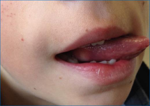
Physical examination revealed a single whitish nodule, firm, slightly tender on palpation, and about 2 cm in size in the postero-lateral right margin of the tongue (Figure 1).

Figure 1 Single whitish nodule, about 2 cm in size in the postero-lateral margin of the tongue. 

As the etiology of the lesion was unknown, and it was a relatively large lesion in a child, an incisional biopsy was done. The histology disclosed a mucosa covered by stratified squamous epithelium exhibiting reactive hyperplasia and a poorly circumscribed and infiltrative lesion on the subepithelial tissue composed of polygonal cells with small nuclei and abundant pale eosinophilic granular cytoplasm (Figure 2A to C). The cell borders were indistinct, giving rise to a syncytial appearance. The mitotic index was low, and there was a strong and diffuse expression of S100, highlighting its putative Schwannian origin. Therefore a diagnosis of granular cell tumor was established. The patient underwent a right partial glossectomy, and the surgical margins were tumor free.

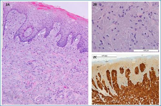
Figures 2A to C (A) H&E 100×: mucosa covered by stratified squamous epithelium exhibiting reactive hyperplasia and a poorly circumscribed and infiltrative lesion on the subepithelial tissue, (B) H&E 400×: it is composed of polygonal cells with small nuclei and abundant pale eosinophilic granular cytoplasm; pustule-ovoid bodies of Millian corresponding to larger granules surrounded by a clear halo, are also identified, (C): strong and diffuse expression of S100 in the neoplastic cells, 200×. 

Discussion

Granular cell tumors (GCT) are rare soft tissue tumors, mostly benign, thought to derive from cells in the nerve sheath1,9. S100 protein is the most widely used immunohistochemical marker for these tumors, which was positive in our case1. Other immunostains positivity has also been described, such as neuron-specific enolase, cluster of differentiation 68, and calretinin4,10,11.

Histologically GCT is characterized by non-encapsulated cords, nests, or sheets of infiltrative polygonal and occasionally spindled cells with an abundant granular eosinophilic cytoplasm. Nuclei are small and centrally placed with dense chromatin4. An important histological finding reported in up to half of granular cell tumors is pseudoepitheliomatous hyperplasia of the overlying epithelium1. Therefore, if the biopsy is too superficial, GCT can be mistaken for a squamous cell carcinoma12.

As GCT generally occurs in adults in the third to sixth decades and is very rare in the first two decades of life, many other benign lesions have to be considered in the differential diagnosis, including vascular lesions, lipoma, fibroma, or mucous cyst13.

Furthermore, multiple GCTs have been rarely reported, especially in children with neurofibromatosis, Noonan’s syndrome, or growth retardation14.

Although benign GCTs have an excellent prognosis after local excision, the malignant ones have a poor prognosis, as they are not sensitive to radiotherapy or chemotherapy. Differentiation is based on histological findings, a complete history, physical examination, and other criteria, including size, rapidity of growth, invasion of nearby structures, and the presence of metastasis. Some authors consider it malignant only if it has metastasized3,8,15. Fanburg-Smith et al. have p roposed six items to consider a granular cell tumor malignant (Table 1)16. If three or more of these criteria are present, then the tumor is considered malignant, in which case it grows faster and has the potential to produce metastasis, especially to the regional lymph nodes, liver, lungs, and bone8.

Table 1 Classification of GCTs according to Fanburg-Smith et al. criteria14  

Criteria
Increased nuclear-to-cytoplasmic ratio
Pleomorphism (celular and/or nuclear)
Tumor necrosis
Spindling of tumor cells
Vesicular nuclei with prominent nucleoli
Mitotic count of > 2 in 10 high-power fields (200× field)
Classification
Benign None of the criteria or focal pleomorphism
Atypical 1-2 criteria
Malignant ≥ 3 criteria

Regardless of the malignancy, election therapy is the simple conservative excision of the lesion4. Relapse occurs more frequently when surgical margins are positive for tumor cells, but some studies have found local relapse even after total excision with free margin17.

This case is interesting because it is an uncommon tumor that rarely presents at such a young age. In conclusion, regardless of pediatric age, a biopsy should not be postponed whenever there is uncertainty in the clinical diagnosis.

REFERENCES

1. Neville BW, Damm DD, Allen CM, Chi AC. Soft tissue tumors. In:Neville BW, Damm DD, Allen CM, Chi AC, eds. Oral and maxillofacial pathology, 4th ed.St Louis:Elsevier, 2016. p.502-3. [ Links ]

2. van de Loo S, Thunnissen E, Postmus P, van der Waal I. Granular cell tumor of the oral cavity;a case series including a case of metachronous occurrence in the tongue and the lung. Med Oral Patol Oral Cir Bucal. 2015;20:e30-3. [ Links ]

3. Mobarki M, Dumollard JM, Dal Col P, Camy F, Peoc'h M, Karpathiou G. Granular cell tumor a study of 42 cases and systemic review of the literature. Pathol Res Pract. 2020;216:152865. [ Links ]

4. Neelon D, Lannan F, Childs J. Granular Cell Tumor. In:StatPearls. Treasure Island (FL):StatPearls Publishing;2021. [ Links ]

5. Muscardin LM, Paradisi M, Provini A, Cota C, Marzetti G. Multiple cutaneous granular cell tumors, joint hypermobility and mild facial dysmorphism in a child. Int J Dermatol. 2006;45:847-50. [ Links ]

6. Marcoval J, Bauer-Alonso A, Llobera-Ris C, Moreno-Vilchez C, Penín RM, Bermejo J. Granular cell tumor:a clinical study of 81 patients. Actas Dermosifiliogr (Engl Ed). 2021;112:441-6. [ Links ]

7. Suchitra G, Tambekar KN, Gopal KP. Abrikossoff's tumor of tongue:report of an uncommon lesion. J Oral Maxillofac Pathol. 2014;18:134-6. [ Links ]

8. Machado I, Cruz J, Lavernia J, Llombart-Bosch A. Solitary, multiple, benign, atypical, or malignant:the “granular cell tumor”puzzle. Virchows Arch. 2016;468:527-38. [ Links ]

9. Musha A, Ogawa M, Yokoo S. Granular cell tumors of the tongue:fibroma or schwannoma. Head Face Med. 2018;14:1. [ Links ]

10. Kurtin PJ, Bonin DM. Immunohistochemical demonstration of the lysosome-associated glycoprotein CD68(KP-1) in granular cell tumors and schwannomas. Hum Pathol. 1994;25:1172e8. [ Links ]

11. Fine SW, Li M. Expression of calretinin and the alpha-subunit of inhibin in granular cell tumors. Am J Clin Pathol. 2003;119:259-64. [ Links ]

12. Ferreira JC, Oton-Leite AF, Guidi R, Mendonça EF. Granular cell tumor mimicking a squamous cell carcinoma of the tongue:a case report. BMC Res Notes. 2017;10:14. [ Links ]

13. Nagaraj PB, Ongole R, Bhujanga-Rao BR. Granular cell tumor of the tongue in a 6-year-old girl - a case report. Med Oral Patol Oral Cir Bucal. 2006;11:E162-4. [ Links ]

14. Tomson N, Abdullah A, Tan CY. Multiple granular cell tumors in a child with growth retardation. Report of a case and review of the literature. Int J Dermatol. 2006;45:1358-61. [ Links ]

15. Pérez-González YC, Pagura L, Llamas-Velasco M, Cortes-Lambea L, Kutzner H, Requena L. Primary cutaneous malignant granular cell tumor:An immunohistochemical study and review of the literature. Am J Dermatopathol. 2015;37:334-40. [ Links ]

16. Fanburg-Smith JC, Meis-Kindblom JM, Fante R, Kindblom LG. Malignant granular cell tumor of soft tissue:diagnostic criteria and clinicopathologic correlation. Am J Surg Pathol. 1998;22:779-94. [ Links ]

17. Moten AS, Movva S, von Mehren M, Wu H, Esnaola NF, Reddy SS, et al. Granular cell tumor experience at a comprehensive cancer center. J Surg Res. 2018;226:1-7. [ Links ]

FundingNone.

Ethical disclosures

Protection of human and animal subjects. The authors declare that no experiments were performed on humans or animals for this study.

Confidentiality of data. The authors declare that they have followed the protocols of their work center on the publication of patient data.

Right to privacy and informed consent. The authors have obtained the written informed consent of the patients or subjects mentioned in the article. The corresponding author is in possession of this document.

Received: December 09, 2022; Accepted: January 18, 2023

*Corresponding author: Pedro R. Matos E-mail: pedro.rolo.matos@chsj.min-saude.pt

Conflicts of interest

None.

Creative Commons License Portuguese Society of Dermatology and Venereology. Published by Permanyer. This is an open access article under the CC BY-NC-ND license