SciELO - Scientific Electronic Library Online

 
vol.81 número3Lúpus eritematoso induzido por fármacosCandidíase cutânea congénita num recém-nascido de termo índice de autoresíndice de assuntosPesquisa de artigos
Home Pagelista alfabética de periódicos  

Serviços Personalizados

Journal

Artigo

Indicadores

Links relacionados

  • Não possue artigos similaresSimilares em SciELO

Compartilhar


Portuguese Journal of Dermatology and Venereology

versão impressa ISSN 2795-501Xversão On-line ISSN 2795-5001

Port J Dermatol Venereol. vol.81 no.3 Lisboa ago. 2023  Epub 08-Mar-2023

https://doi.org/10.24875/pjdv.23000004 

CASE REPORT

Keratosis lichenoides chronica: a diagnosis to remember

Queratose liquenóide crónica: um diagnóstico a relembrar

Ana S. Pereira1  *  https://orcid.org/0000-0003-4930-4614

José C. Cardoso1 

Mariana Batista1 

1Department of Dermatology, Centro Hospitalar e Universitário de Coimbra, Coimbra, Portugal


Abstract

Keratosis lichenoides chronica (KLC) or Nekam's disease, is an uncommon dermatosis, assumed for years as a variant of another inflammatory dermatosis (such as cutaneous lupus erythematosus or lichen planus) but currently accepted as a distinctive condition. Although pathophysiologic mechanisms need further research, clinical aspects of KLC are well-characterised, particularly lichenoid hyperkeratotic papules arranged linearly or in a reticulate pattern over the extremities, seborrheic-like dermatitis on the face and oral or genital erosions. Histopathology usually shows lichenoid interface dermatitis with numerous necrotic keratinocytes and parakeratosis. Keratosis lichenoides chronica (KLC) typically has a chronic progressive course with poor response to treatment. In the following case, we present a 56-year-old man with chronic dermatosis whose clinicopathological findings allowed the diagnosis of KLC. The patient was treated with acitretin, topical steroids and topical calcineurin inhibitors with partial improvement of the lesions.

Keywords Interface dermatitis; Keratosis lichenoides chronica; Lichenoid eruptions; Nekam's disease

Resumo

A Queratose liquenóide crónica (QLC) ou Doença de Nekam é uma dermatose pouco comum, entendida desde há anos como variante de outra dermatose inflamatória (como lúpus eritematoso cutâneo ou líquen plano), mas atualmente aceite como uma entidade distinta. Embora os seus mecanismos fisiopatológicos necessitem de mais investigação, os aspetos clínicos da QLC encontram-se bem caracterizados, particularmente pelas pápulas queratósicas liquenóides dispostas linearmente ou num padrão reticulado sobre as extremidades, dermatite seborreica facial e erosões orais ou genitais. A histopatologia demonstra geralmente uma dermatite de interface líquenóide associada a numerosos queratinócitos necróticos e cobertos por paraqueratose. A QLC tem tipicamente um curso progressivo crónico com resposta insatisfatória ao tratamento. No caso seguinte, apresentamos um homem de 56 anos de idade com uma dermatose crónica, cujos achados clinicopatológicos permitiram o diagnóstico de QLC. O doente foi tratado com acitretina, esteróides tópicos e inibidores de calcineurina tópicos com melhoria parcial das lesões.

Palavras-chave Dermatite de interface; Doença de Nekam; Erupções liquenóides; Queratose liquenóide crónica

Introduction

Keratosis lichenoides chronica (KLC) is an uncommon mucocutaneous disorder originally described in 1895 by Kaposi, who called it ‘lichen ruber acuminatus morbilliform’1. Later, in 1938, Nekam took note of acrosyringeal hyperkeratosis in the case published by Kaposi and named the disease ‘porokeratosis striata lichenoides’ despite the lack of coronoid lamella2. Finally, in 1972, Margolis et al. introduced the nomenclature currently used—‘KLC’3.

Case description

A 56-year-old male, otherwise healthy, presented to the dermatology department with a 1-year history of persistent asymptomatic cutaneous lesions predominantly affecting the limbs and face. On examination, multiple keratotic violaceous papules and plaques were distributed over the dorsum of his hands and fingers (Fig. 1A), as well as on the medial aspect of both thighs, where they were arranged in a linear pattern (Fig. 1B). On his face, there were erythematous and squamous lesions located mainly in the midfacial area, resembling seborrheic dermatitis (Fig. 1C). Mucous membranes, genital region, scalp and nails were spared. The remaining physical examination was unremarkable and his routine blood tests, including viral serologies and autoimmunity markers, were normal.

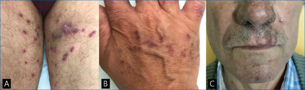
Figure 1 Clinical features. A: keratotic papules on the thighs, some arranged in a linear pattern; B: keratotic violaceous papules over the dorsum of the hand; C: facial seborrheic dermatitis-like eruption. 

There was no familiar history of similar complaints.

A skin biopsy performed on a thigh lesion revealed irregular acanthosis and hyperkeratosis with alternating areas of orthokeratosis and parakeratosis. In the upper dermis, there was a dense lichenoid infiltrate composed of lymphocytes and plasma cells, associated with multiple foci of vacuolar degeneration of the basal epidermal layer and apoptotic keratinocytes (Fig. 2).

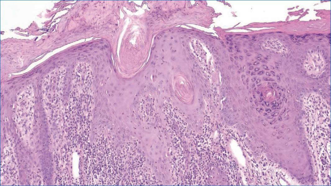
Figure 2 Histopathological aspects: irregular acanthosis with focal parakeratosis, apoptotic keratinocytes at various levels of the epidermis and mixed lichenoid infiltrate in the superficial dermis (H&E, 10x). 

This combination of clinical and histological findings strongly suggested the diagnosis of KLC. The patient declined to be tested for the nucleotide-binding domain and leucine-rich repeat-containing protein 1 (NLRP1) gene. Treatment was initiated with acitretin 35 mg daily, in addition to the topical application of tacrolimus 1% ointment on the face and clobetasol propionate ointment 0.05% on the extremities. At 6 months follow-up, the lesions on his hands had significantly improved. However, other lesions showed milder responses to the treatment.

Discussion

Keratosis lichenoides chronica (KLC), also known as Nekam's disease, is a rare dermatosis of uncertain etiology. It appears most frequently in adults, with a peak of incidence in the 4th decade and slight predominance in males1,2,4.

Previously perceived as the expression of other inflammatory conditions such as lichen planus, lupus erythematosus or lichen simplex chronicus5,6, KLC is now considered a distinct entity since the detection of a gain-of-function mutation in the nucleotide-binding domain and leucine-rich repeat-containing proteins 1 (NLRP1) gene in a family with semi-dominantly inherited KLC7.

NLRP1 is an inflammasome sensor protein in high levels in keratinocytes and cutaneous fibroblasts. It is thought that the gain-of-function mutation in the NLRP1 gene causes constitutive inflammasome activation and leads to reactive keratinocyte proliferation7,8. However, further studies are required to fully comprehend the pathogenesis of both familiar and sporadic cases.

Clinically, KLC is characterised by lichenoid or keratotic violaceus papules arranged in a linear or reticulated pattern, especially on the limbs4. A seborrheic-like dermatitis eruption on the face is also a common finding of this disorder4. Less frequent features include oral and genital ulcerative lesions, palmoplantar keratoderma and onychodystrophy which are found, respectively, in 50, 40 and 30% of cases1,9,10.

Regarding histology, typical findings are hyperkeratosis with focal parakeratosis, irregular acanthosis alternating with areas of atrophy, vacuolar degeneration of keratinocytes at dermoepidermal junction and chronic inflammatory infiltrate in the upper dermis consisting of lymphocytes, histiocytes and plasma cells (often around infundibula and acrosyringia)4,5.

Several inflammatory dermatoses, such as lichen planus, lichen planus-psoriasis overlap, lichen planopilaris, pityriasis rubra pilaris and cutaneous lupus erythematous may mimic KLC10.

Keratosis lichenoides chronica (KLC) has a chronic and progressive course and is highly resistant to therapy11,12. The most effective treatment consists of oral retinoids at a dose of 0.3-0.6 mg/kg/day2,11, although complete responses are infrequent11. Phototherapy (psoralen plus ultraviolet A and narrow-band ultraviolet B) can be helpful, especially as an adjuvant treatment4,11. Topical treatment, as well as systemic steroids, other immunosuppressants and antimalarials, are usually ineffective2,12.

This case serves to highlight KLC as a well-defined disease with consistent clinical and histological characteristics. Its pathogenesis is still not fully understood, although recent progress has been made with the discovery of the possible contribution of the NLRP1 gene.

Despite its rarity, KLC is an entity that dermatologists should keep in mind, with the aim of detecting more cases and performing more investigations on both pathophysiology and therapeutical approaches.

REFERENCES

1. Wozniacka A, Schwartz RA, Omulecki A, Lesiak A, Sysa-Jedrzejowska A. Keratosis lichenoides chronica a diagnostic and therapeutic challenge. Clin Exp Dermatol. 2006;31:48-50. [ Links ]

2. Martins L, Junior D, Almeida V, Horne M, Follador I. Keratosis lichenoides chronica - Case report. An Bras Dermatol. 2011;86:S148-51. [ Links ]

3. Margolis MH, Cooper GA, Johnson S. Keratosis lichenoides chronica. Arch Dermatol. 1972;105:739-43. [ Links ]

4. Aromolo IF, Giacalone S, Genovese G, Maronese CA, Marzano AV. Keratosis lichenoides chronica:a case report and focused overview of the literature. Australas J Dermatol. 2022;63:e99-2. [ Links ]

5. Boer A. Keratosis lichenoides chronica:proposal of a concept. Am J Dermatopathol. 2006;28:260-75. [ Links ]

6. Tchernev G, Nenoff P. Antigen mimicry followed by epitope spreading:a pathogenetic trigger for the clinical morphology of lichen planus and its transition to graham lassueur piccardi little syndrome and keratosis lichenoides chronica - medical hypotheses or reality?An Bras Dermatol. 2009;84:682-8. [ Links ]

7. Zhong FL, Mamai O, Sborgi L, Boussofara L, Hopkins R, Robinson K, et al. Germline NLRP1 mutations cause skin inflammatory and cancer susceptibility syndromes via inflammasome activation Cell. 2016;67:187-202.e17. [ Links ]

8. Konstatinov KN, Søndergaard J, Izuno G, Obreshkova E. Keratosis lichenoides Chronica. J Am Acad Dermatol. 1998;38:306-9. [ Links ]

9. Demirci E, Boyvat A, Arica IE, Kocyigit P, Ozdemir E, Heper AO. Keratosis lichenoides chronica:marked response to PUVA in combination with acitretin. Acta Derm Venereol. 2005;85:552-3. [ Links ]

10. Aruna C, Ramamurthy D, Neelima T, Bandaru H. Nekam's disease. Indian Dermatol Online J. 2016;7:520-2. [ Links ]

11. Adişen E, Erdem O, Celepçi S, Gürer MA. Easy to diagnose, difficult to treat:keratosis lichenoides chronica. Clin Exp Dermatol. 2010;35:47-50. [ Links ]

12. Pistoni F, Peroni A, Colato C, Schena D, Girolomoni G. Keratosis lichenoides chronica:case-based review of treatment options. J Dermatol Treat. 2016;27:383-8. [ Links ]

FundingNone.

Ethical disclosures

Protection of human and animal subjects. The authors declare that no experiments were performed on humans or animals for this study.

Confidentiality of data. The authors declare that they have followed the protocols of their work center on the publication of patient data.

Right to privacy and informed consent. The authors have obtained the written informed consent of the patients or subjects mentioned in the article. The corresponding author is in possession of this document

Received: January 17, 2023; Accepted: February 21, 2023

*Correspondence: Ana S. Pereira E-mail: sfigpereira@gmail.com

Conflicts of interest

None.

Creative Commons License Portuguese Society of Dermatology and Venereology. Published by Permanyer. This is an open access article under the CC BY-NC-ND license